
 Almost two years ago we took Max's lower jaw to California Imaging and Diagnostics for a CT scan. We got some fascinating data that raised a lot of new questions, so to help answer them we recently took a second mastodon to CID for scanning.The particular specimen is from the San Diego Canal, and includes a nearly complete lower jaw and a partial cranium. The cranium was of particular interest to us. It was broken in such a way as to include the entire left tooth row including the tusk, but the fragment was narrow enough to fit through the medical CT scanner; a complete adult mastodon skull is typically far too large to fit through the scanner. Based on tooth wear, while this mastodon was an adult he (it appears to be a male) was as much as 10 years younger than Max, so it gives us an interesting point for comparison. We're still examining the data, so what I'm posting here is preliminary (I only examined most of the data myself for the first time yesterday):Here's a transverse section through the SDC specimen's lower jaw, cutting through the last roots on the 3rd molars:
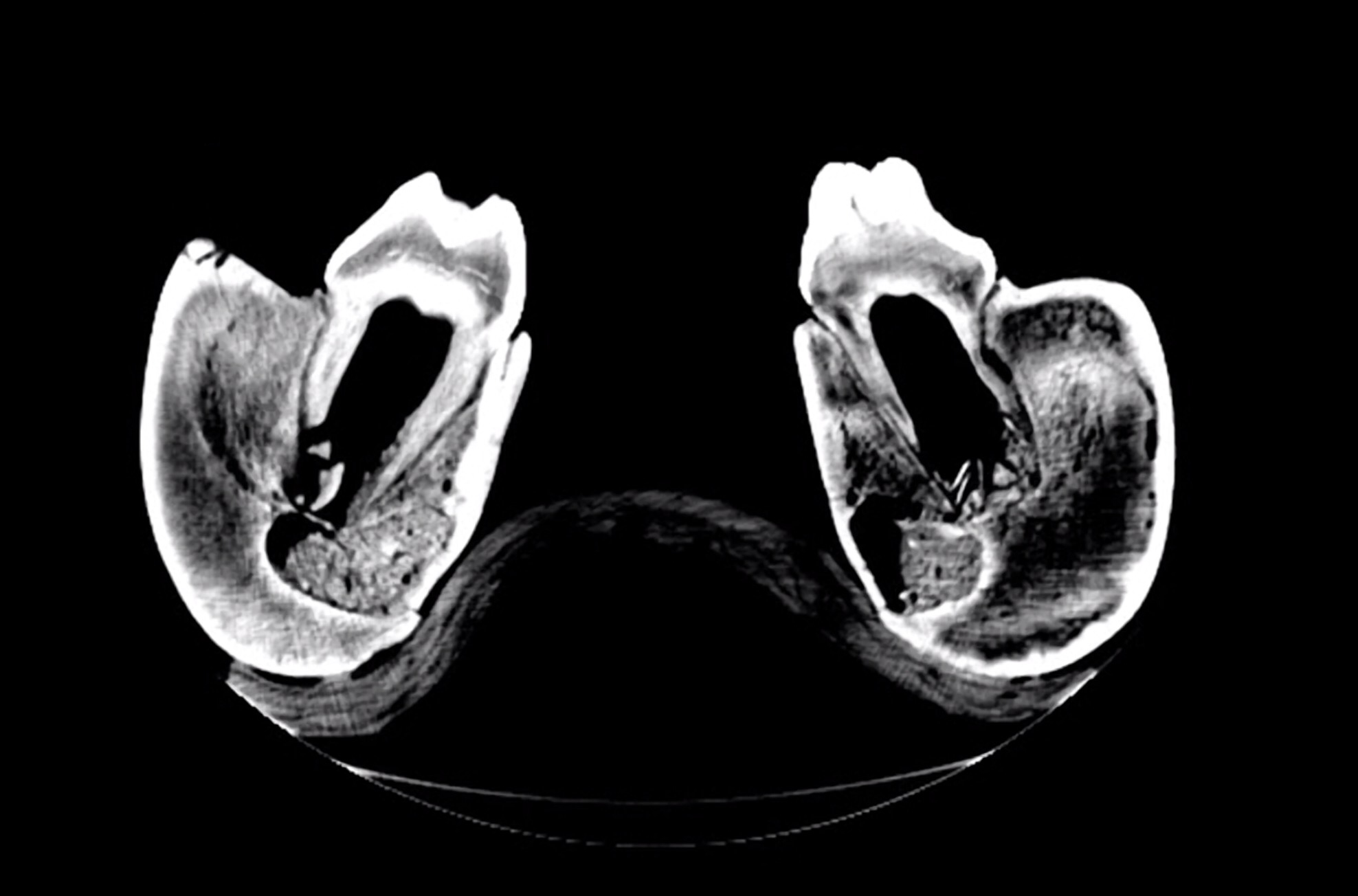 For comparison here's a similar section through Max's jaw:
 The SDC mastodon was a young animal, probably in his early 20's. His 3rd molars were still growing, and had large open pulp cavities and short roots. Max was a middle-aged mastodon in his mid-to-late 30's, with fully developed 3rd molars with deep roots and nearly closed pulp cavities.Here's a sagittal section through the SDC specimen's right dentary, followed by a similar section on Max:
 This view also emphasizes the huge open pulp cavity in the SDC specimen compared to Max's nearly solid teeth.Here's another sagittal section through the SDC right dentary, on a slightly different plane than the image above:
 Notice the differently-textured bone just in front of the 2nd molar, outlined in red below:
 With their horizontal tooth replacement, proboscidean teeth are constantly moving forward in the jaw, which means the bone in the jaw is constantly being absorbed and redeposited. The SDC specimen had recently lost his first molar, and the socket is filled by young, spongy bone that shows up as a different texture in the scan. In the much older Max this part of the jaw is as dense as the surrounding bone.Below is a transverse section through the cranium, followed by a marked-up version. I've rotated the image so that it's approximately in life orientation (it was upside down in the scanner):
 The blue area is the 3rd molar, at about the level of the 2nd lophid. The red area is actually the bottom of the unerupted part of the tusk, which extends all the way back at least to the 3rd molar. We can't tell in this specimen just how far back the tusk goes, because it's curving upward and the upper part of the cranium wasn't preserved.Finally, here's a section though a more distal part of the tusk:
 Again, this is rotated into approximately life orientation. One side of the tusk is damaged, and splinters of the tusk have broken off the inside and are laying haphazardly in the pulp cavity (which also contains some sediment). The better-preserved area shows visible concentric lines, the tusk's growth rings.We'll be studying these new scans in detail over the summer and fall, so there will be more to come. Thanks to CID for providing access to their CT scanner and running these scans for us!