
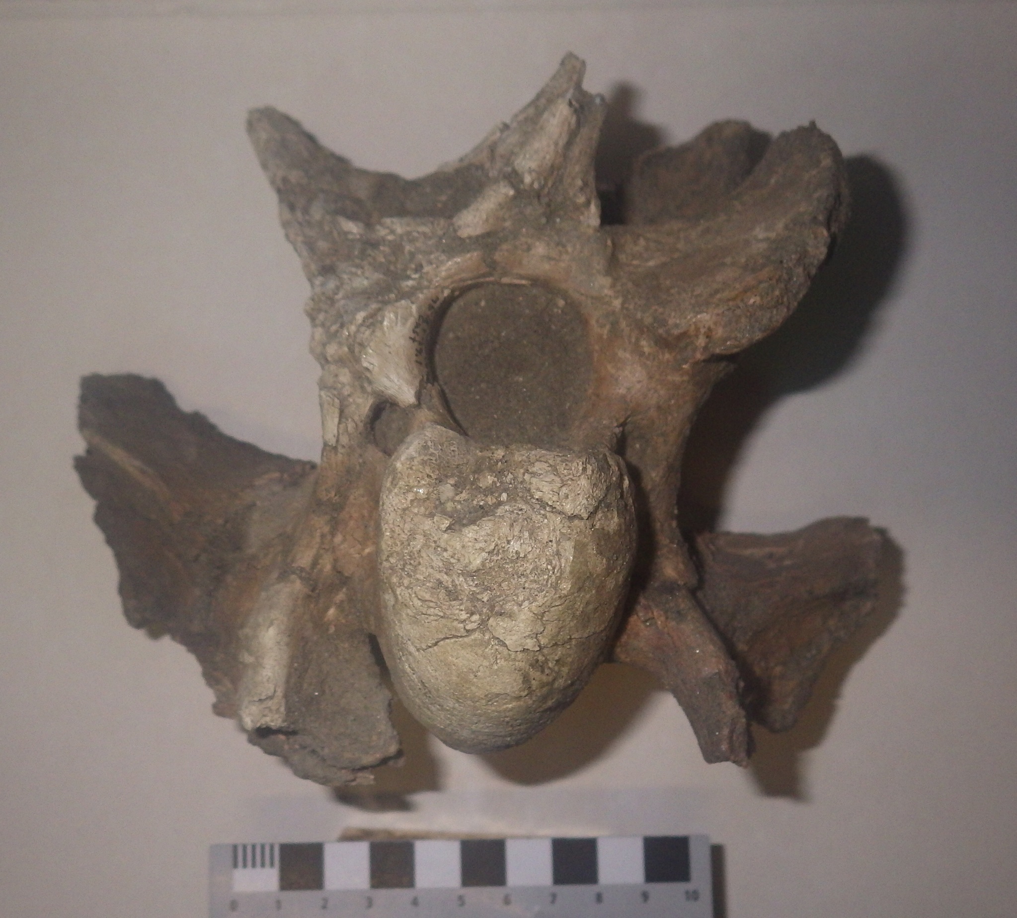
 For today's Fossil Friday we have a vertebra from one of the more common Pleistocene animals in the region, the bison.The vertebral column (backbone) in mammals is typically divided into five separate regions, based on common characteristics and the location in the body. From front to back these are the cervical vertebrae, which form the neck; thoracic vertebrae, which make up part of the back and have ribs attached; the lumbar vertebrae, which form the lower back; sacral vertebrae, which form part of the hip girdle; and caudal vertebrae, which make up the tail.Nearly all mammals have vertebrae from each of these regions. The main exceptions are whales and sea cows, which have no sacral vertebrae. (In fact, they almost certainly do have sacrals, but they are modified to the point that they're indistinguishable from lumbar vertebrae. Very primitive whales and sea cows still had identifiable sacrals.) Even tailless mammals such as humans still have caudal vertebrae; our tailbone, or coccyx, is formed by five fused caudal vertebrae.Another peculiarity of mammals is that almost every living species has exactly 7 cervical vertebrae, whether the neck is short, long, rigid, or flexible. In many animals the cervical vertebrae are very intricate, with all kinds of slender projections and holes passing through them. This happens because a lot of things are happening in the neck. Besides the spinal cord passing through the vertebrae (as it does in all vertebrae) the carotid arteries and jugular veins run alongside the cervicals as they carry blood to and from the brain, both the esophagus and trachea pass directly under the cervicals, and there are numerous muscles and their associated nerves and tendons that control the complex movement of the head and neck. With so much going on, these vertebrae have to be complex to make room for everything while still providing attachment areas for all the neck muscles.The image at the top is an anterior (or cranial) view of a bison cervical vertebra. The bone should be close to symmetrical, but has been somewhat deformed, probably after burial. The prominent oval structure near the bottom is the anterior articulation, the ball part of a "ball-and-socket" joint with the next vertebra forward. Immediately above that is a dark circular area (dark because it's still filled with sediment) called the neural canal, which is the passage for the spinal cord.Here's a posterior (caudal) view of the same vertebra:
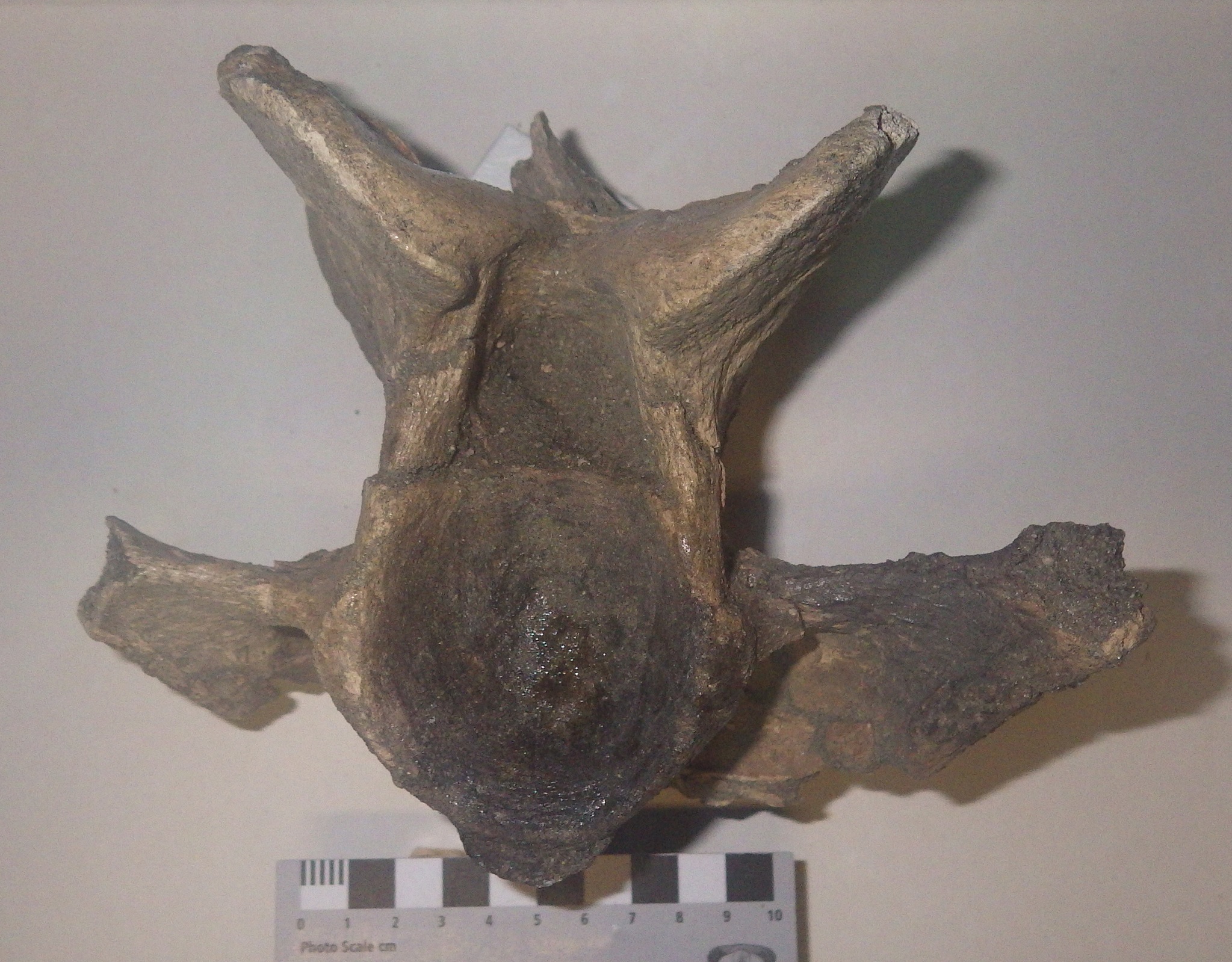 The round area at the bottom is the posterior articulation, which is a concave socket that articulates with the ball from the next posterior vertebra. The sediment-filled neural canal is the bell-shaped structure immediately above the articulation.This is the left lateral view, with anterior to the left:
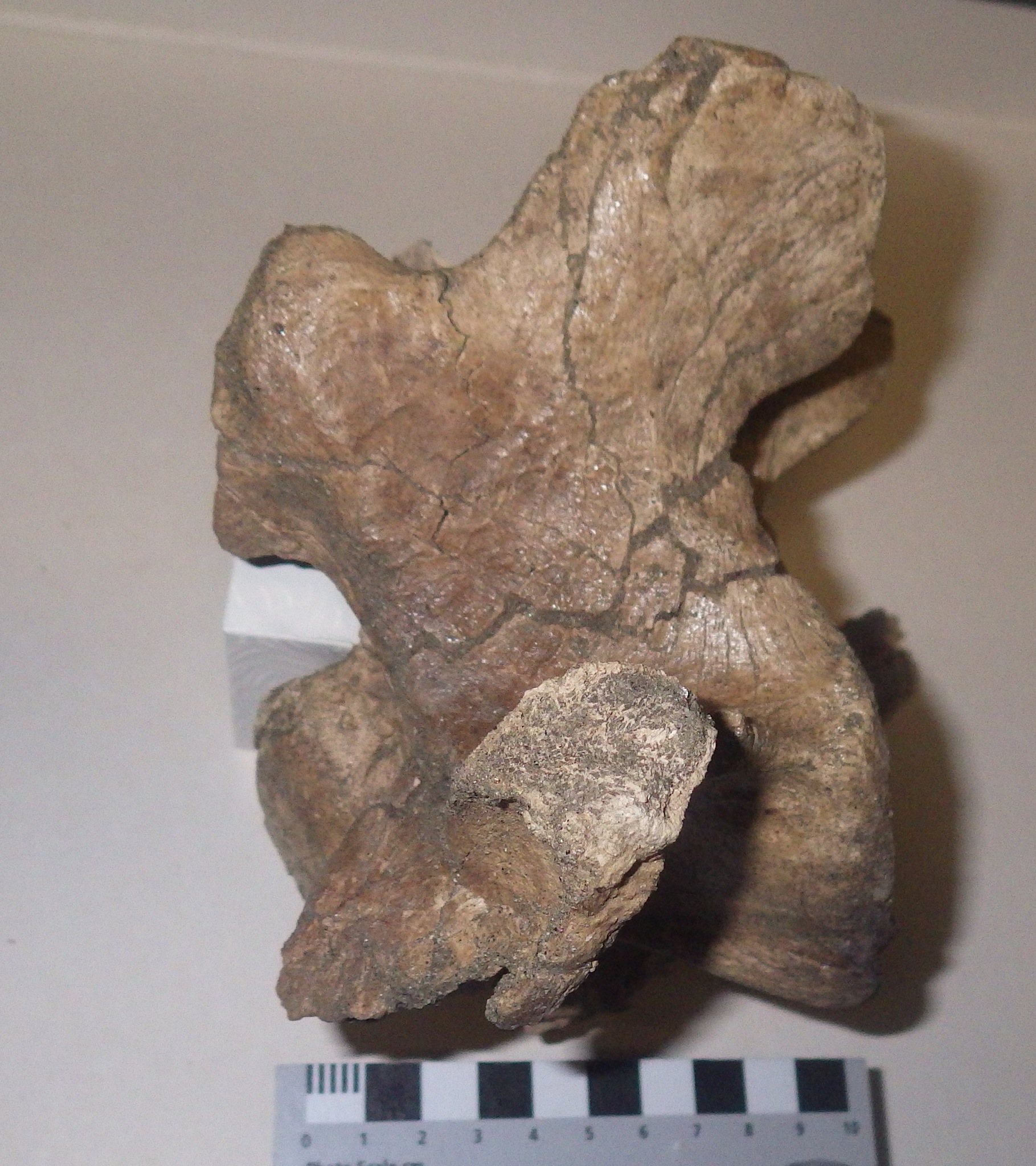 The vertebra should actually be quite a bit taller, but most of the neural spine is missing. The neural spine is a prominent bony projection along the top of the vertebrae that serves as an attachment point for some of the muscles responsible for raising the head.The label on this specimen identifies it as the fourth cervical vertebra, which looks like a pretty good match to me (compare with the modern bison vertebrae shown here). There are two different species of bison known from Southern California, the long-horned Bison latifrons and the (relatively) short-horned Bison antiquus. Based on its size this vertebra probably came from the smaller B. antiquus.This vertebra was collected close to the same locality as the mastodon tooth from last week's Fossil Friday, about 15 miles southwest of the museum.