

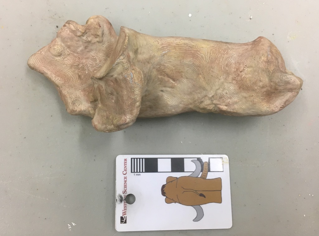
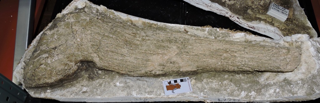
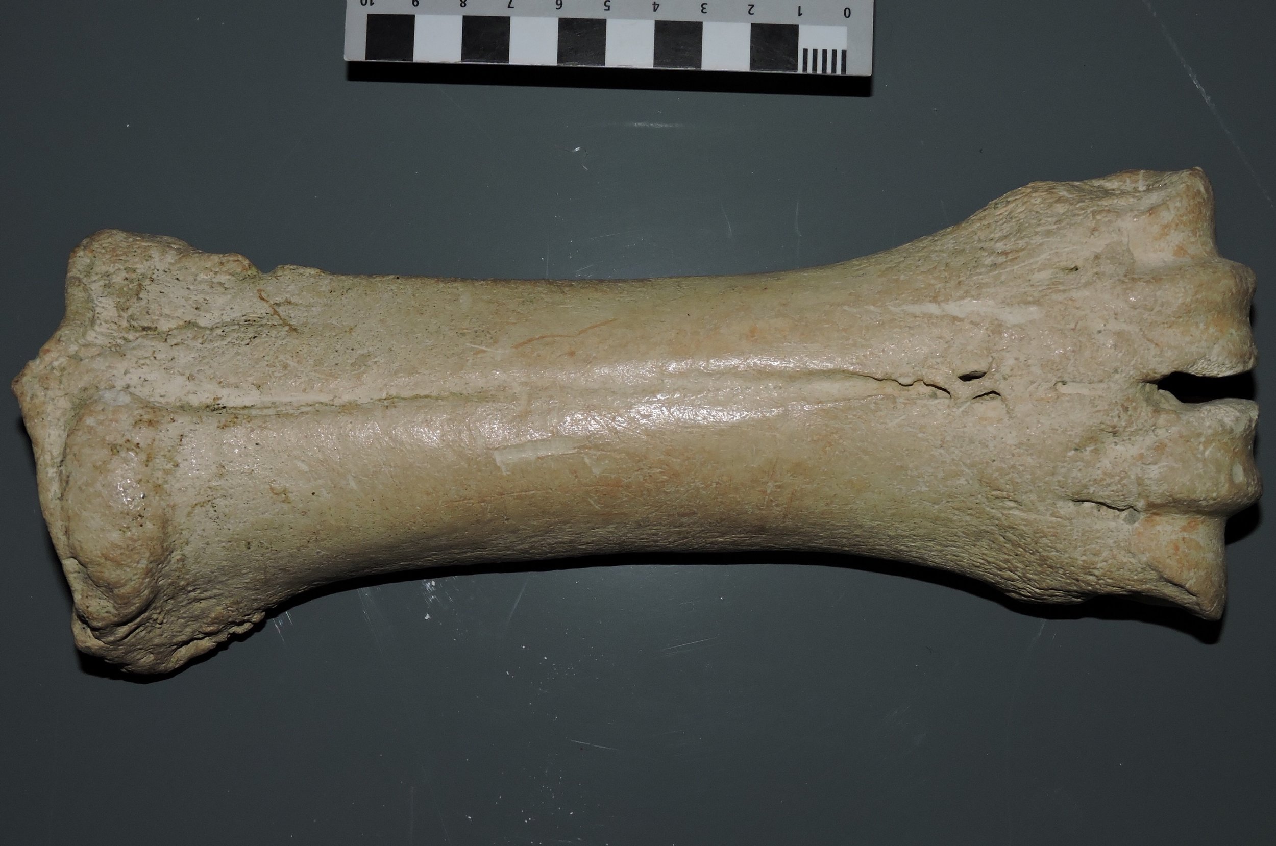
 At our annual Science Under the Stars fundraiser last September, or donors provided us with funding to start a #D scanning and printing lab the the Western Science Center. While some of our equipment is still on order, our first printer arrived a few weeks ago and we've been printing as much as possible while we train ourselves in its use. During the Valley of the Mastodons Symposium, Bernard Means from the Virtual Curation Lab scanned a number of our specimens. While we wait for our scanner to arrive we've been printing some of Bernard's scans.Earlier this week we printed a right Bison calcaneum, seen above in medial view. The original specimen is exhibited in the floor case with the mastodon "Little Stevie" (they were found together). It's difficult to photograph through this case (below), and it's difficult to open; the symposium is the first time it has been opened in 11 years! So while the case was opened we scanned as much as possible, including this calcaneum (which is visible in the center of the case photo below):
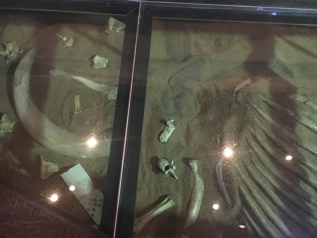 This bone was our largest single print to date, and took about 24 hours to print. We've since printed a slightly larger bone as we continue to explore the limits of the printer.
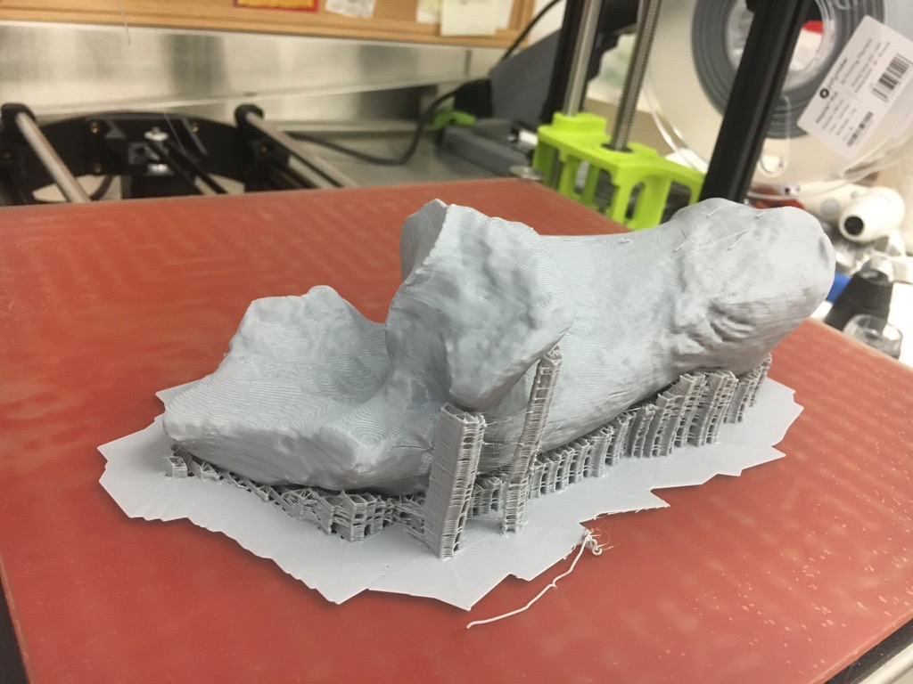 The calcaneum is part of the foot and ankle structure, and forms the heel and the attachment point for the Achilles tendon. This particular specimen was collected at Diamond Valley Lake's East Dam, and is roughly 45,000 years old. The original specimen is now back on display in its floor case, while the printed replica will be added to our teaching collection.
Fossil Friday - Bison molar
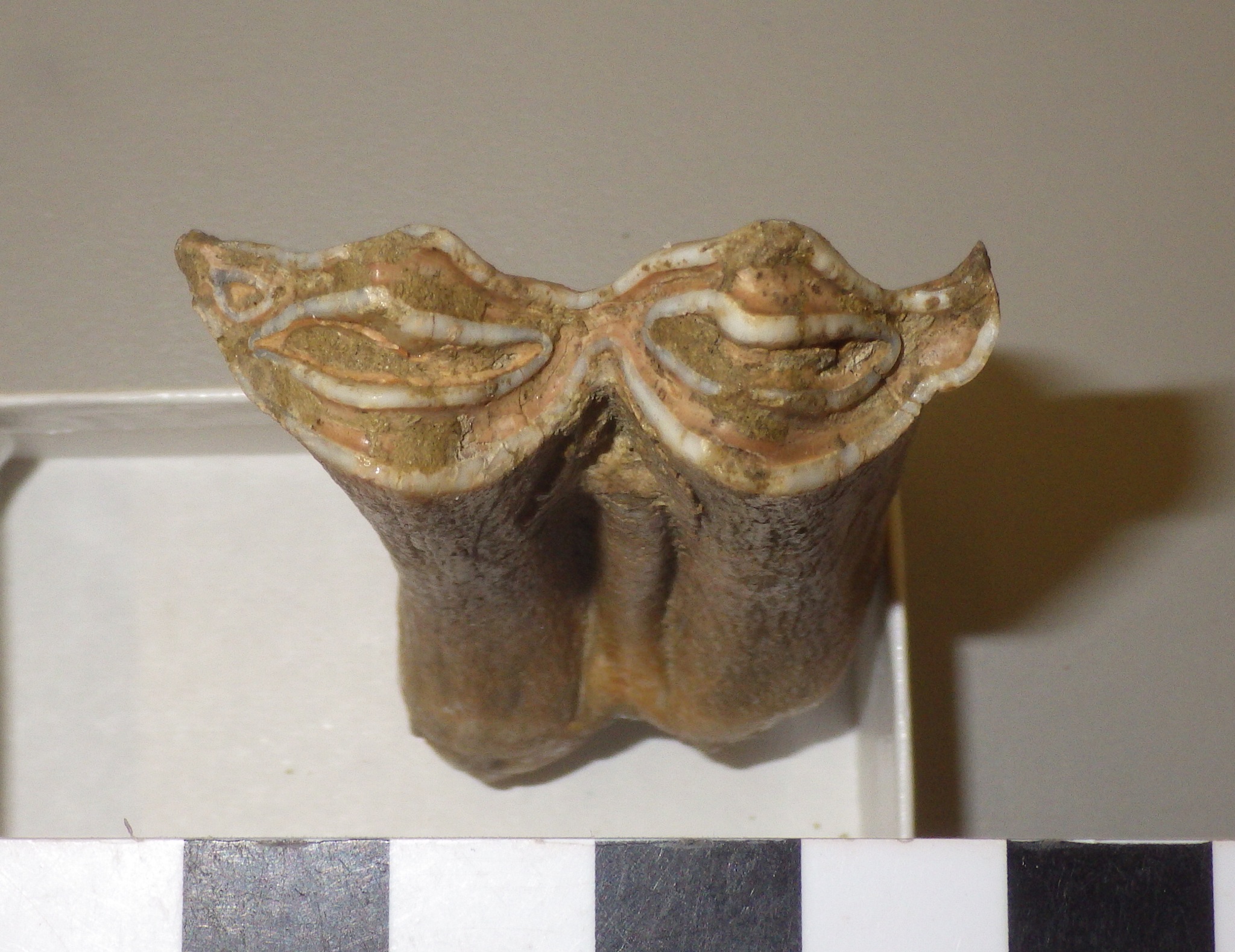
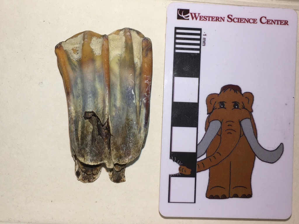 At Diamond Valley Lake, five genera account for 93% of the preserved large animals: Bison, Equus, Camelops, Mammoth, and Paramylodon. Of these five, the most abundant are Bison.The tooth shown above is an upper left 1st molar from a bison. Unfortunately, I photographed it upside down, so even though it's an upper tooth in the image the edge at the top is the chewing surface and the roots are at the bottom. This is the labial view, the side of the tooth closest to the lips.Below is the lingual view, showing the side of the tooth closest to the tongue:
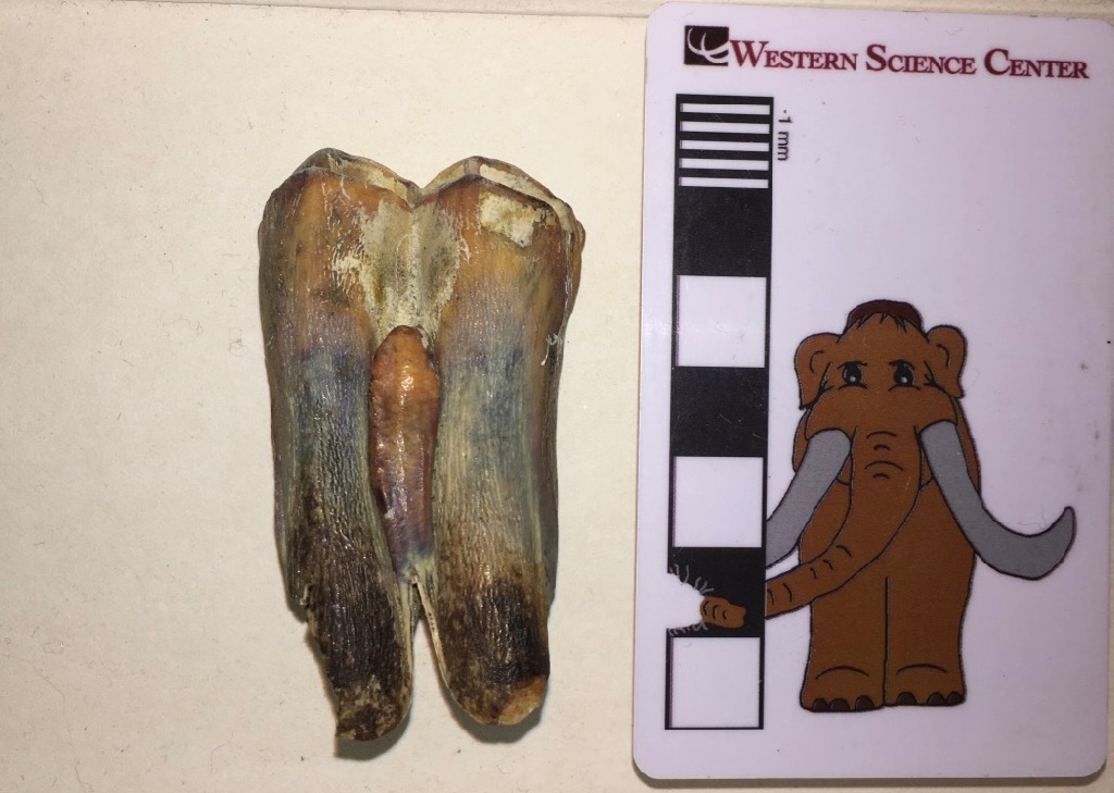 Again, the occlusal edge is at the top, roots at the bottom. The reddish-brown protuberance in the middle of the tooth is a column called the style (stylid on the lower teeth). The style is almost always present in the upper and lower molars of Bison, but it's very rare in the closely related cattle genus Bos. So it's a very convenient identification feature (if not necessarily 100% reliable) for distinguishing between Bos and Bison.In occlusal view (below), the style can be seen end-on on the lingual side of the tooth (on the left in this image):
 This particular tooth only has very light wear. In a heavily-worn molar, the tooth eventually wears away so much that the occlusal surface of the tooth is level with the tip of the style. The style then begins to wear as well, and becomes visible on the occlusal surface as an isolated ring of enamel. Because the style is not the same shape throughout its length, as wear continues the ring gradually becomes an oval, and then a loop of enamel connected to the rest of the tooth. So the style shape on the occlusal surface give a rapid indication of how worn the tooth is, even is it's still embedded in the socket.This tooth was collected from the West Dam of Diamond Valley Lake. Bison latifrons has not been identified from the West Dam, so this is most likely Bison antiquus. Western Science Center also has teaching kits based on interpretation of bison tooth wear in cast specimens available for purchase.
Fossil Friday - Bison atlas vertebra, part 2
 This week's Fossil Friday features one of Diamond Valley Lake's most common large mammals, the bison.Specifically, this bone is the atlas vertebra, the first vertebra in the neck. It's shown above in anterior view. The large hole in the bone is the neural canal, which carries the spinal cord in the live animal. The large concavities on each side of the neural canal are the articulations for the skull's occipital condyles.Below is the posterior view:
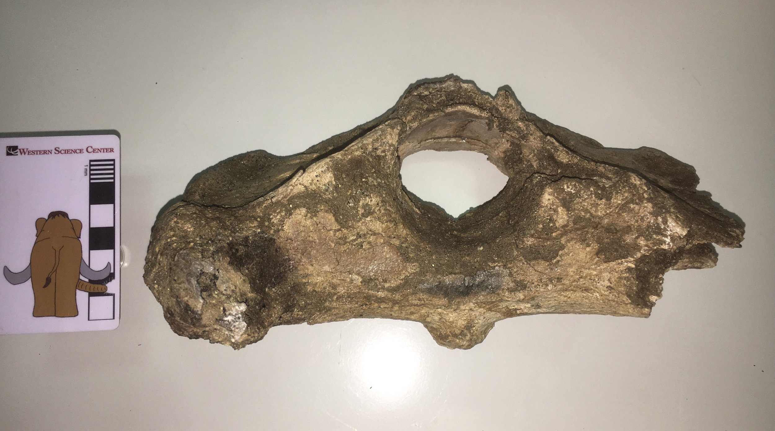 The preservation is a little rougher on this side. The posterior edge of the neural arch (the top of the neural canal) is missing, as is most of the right transverse process. The flat areas to each side of and slightly below the neural canal are the articular surfaces for the 2nd cervical vertebra, the axis.Here is the dorsal view:
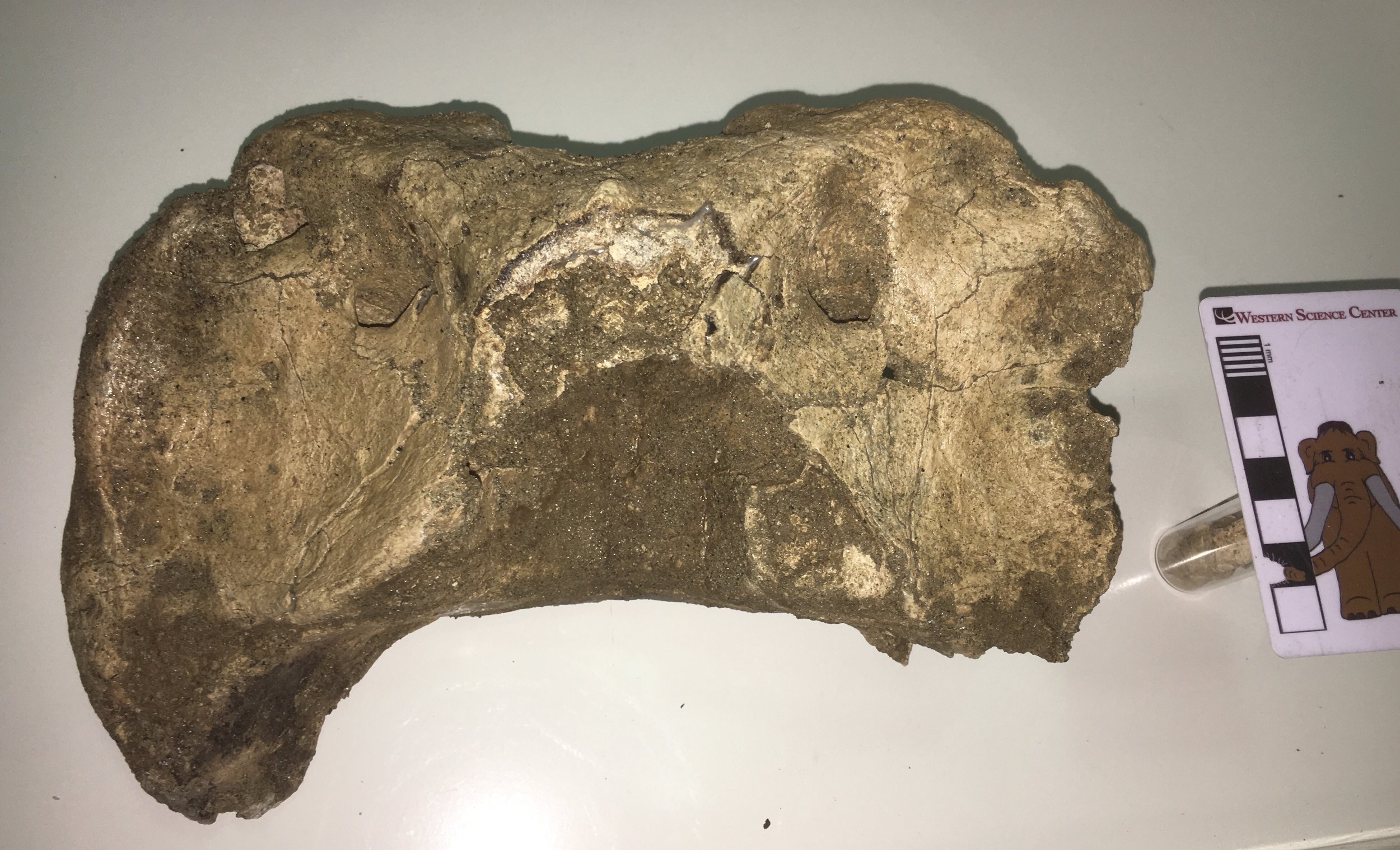 ...and the ventral view:
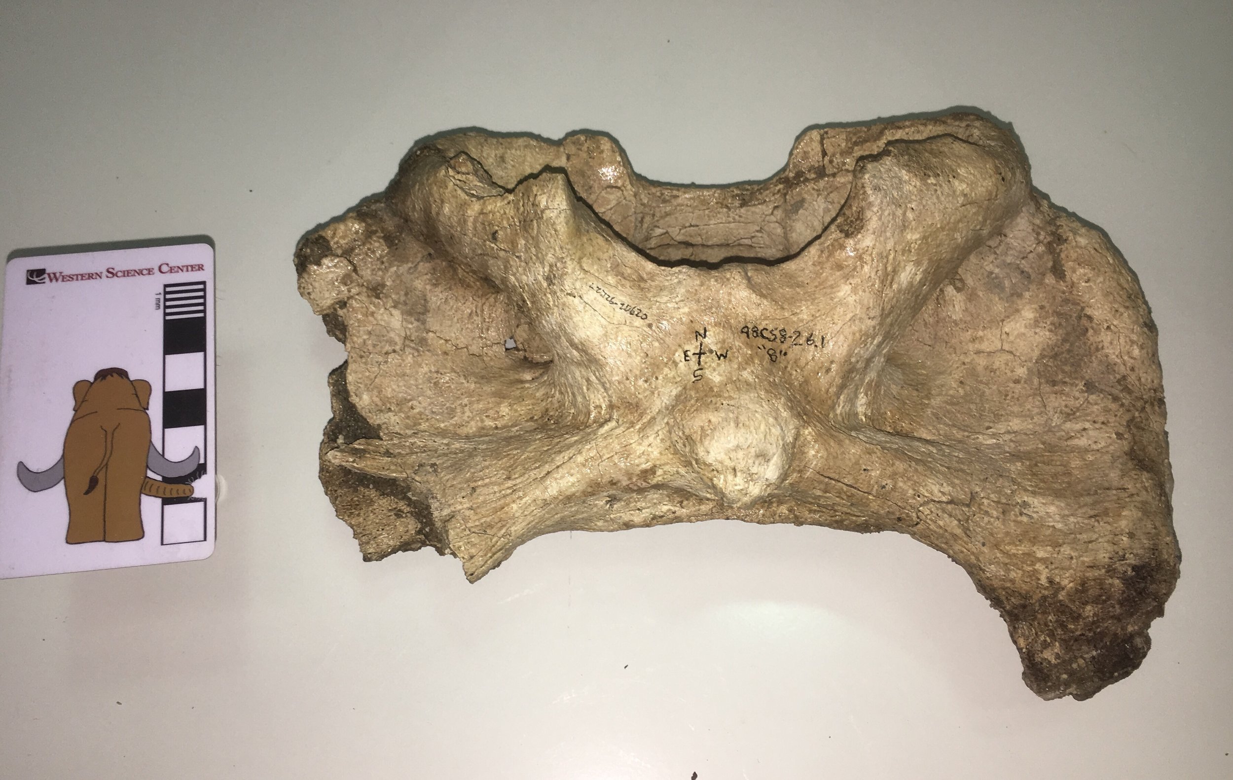 This vertebra is identified in our records as Bison antiquus. It comes from the West Dam area of the lake; sediments there are mostly less than 20,000 years old and seem to only produce Bison antiquus and not the large Bison latifrons. Bison antiquus is generally considered to be closely related or ancestral to the modern Bison bison, and indeed this atlas looks very similar to a modern bison's. This make an interesting contrast to the DVL bison atlas I wrote about back in May 2015. That vertebra was identified as Bison sp., but its shape is a bit different from this one (I even commented at the time that it differed from Bison bison). So perhaps our two extinct bison species really do have distinct atlas shapes. At some point we'll have to compare these bones to atlases that are associated with skulls to confirm that we're seeing specific differences and not individual or sexual variation.
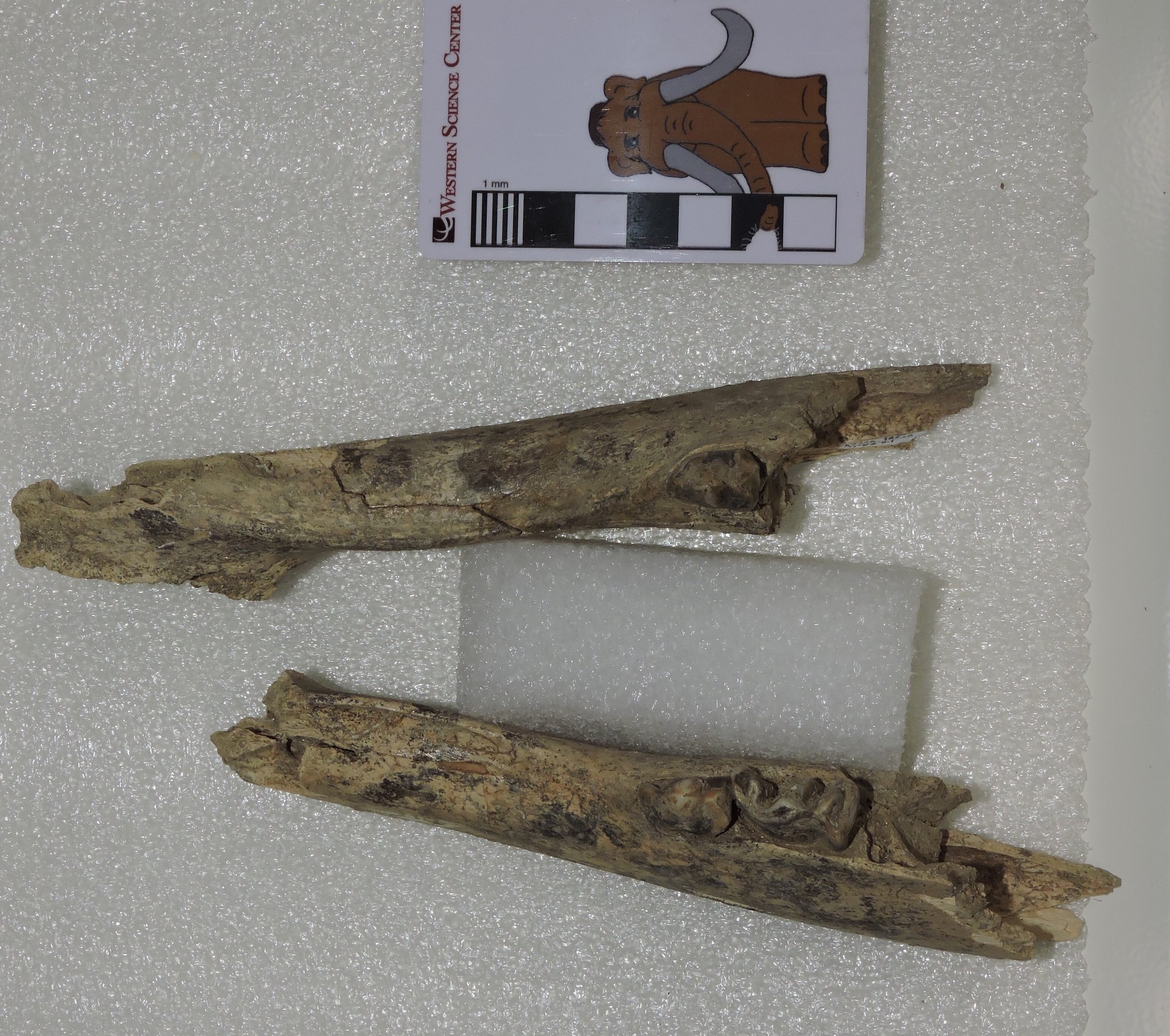
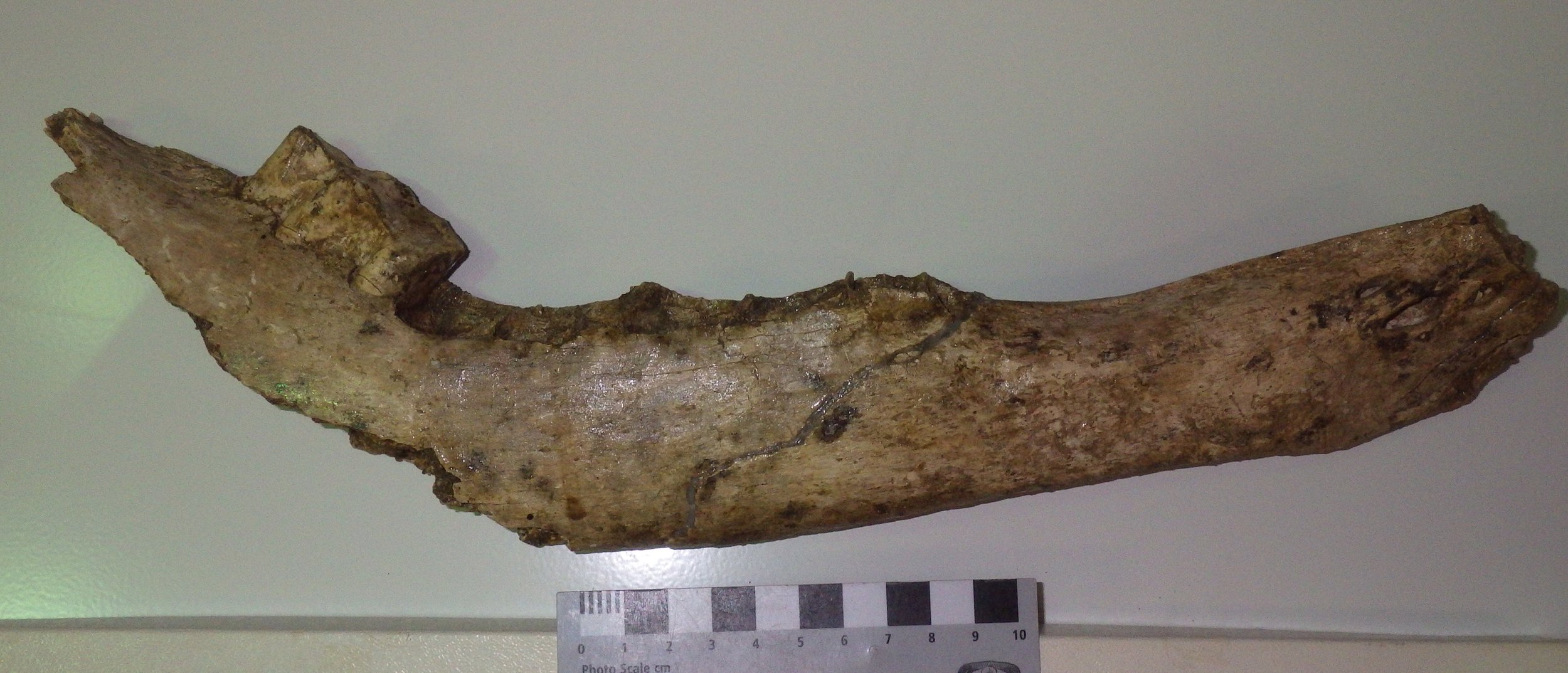
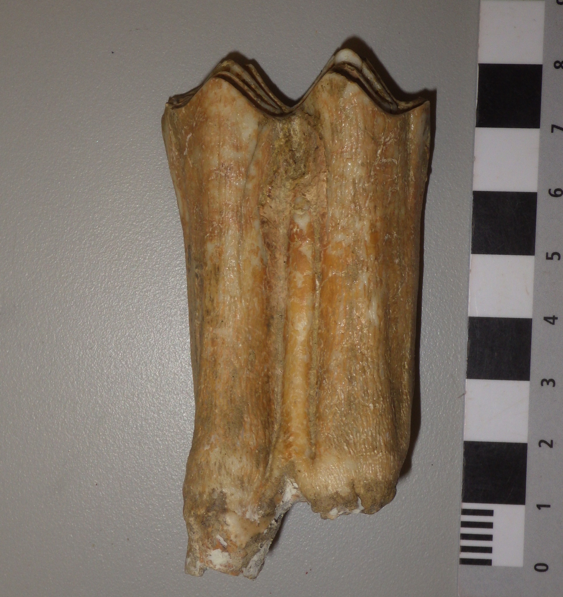
Fossil Friday - chewed-up Bison tibia
 I recently finished reading Anthony Martin's book about dinosaur trace fossils, Dinosaurs Without Bones, so I've had trace fossils on my mind. Even though I'm not a trace fossil specialist I find them intriguing, because they are essentially fossilized behavior. The bone shown here is the distal part of a right tibia from a bison (it's from the older end of the valley, so it could be either Bison antiquus or Bison latifrons). Above is the anterior view, with the bottom of the bone (at the ankle joint) on the left. The proximal part, including the knee joint, is missing. Below is the posterior view of the same bone:
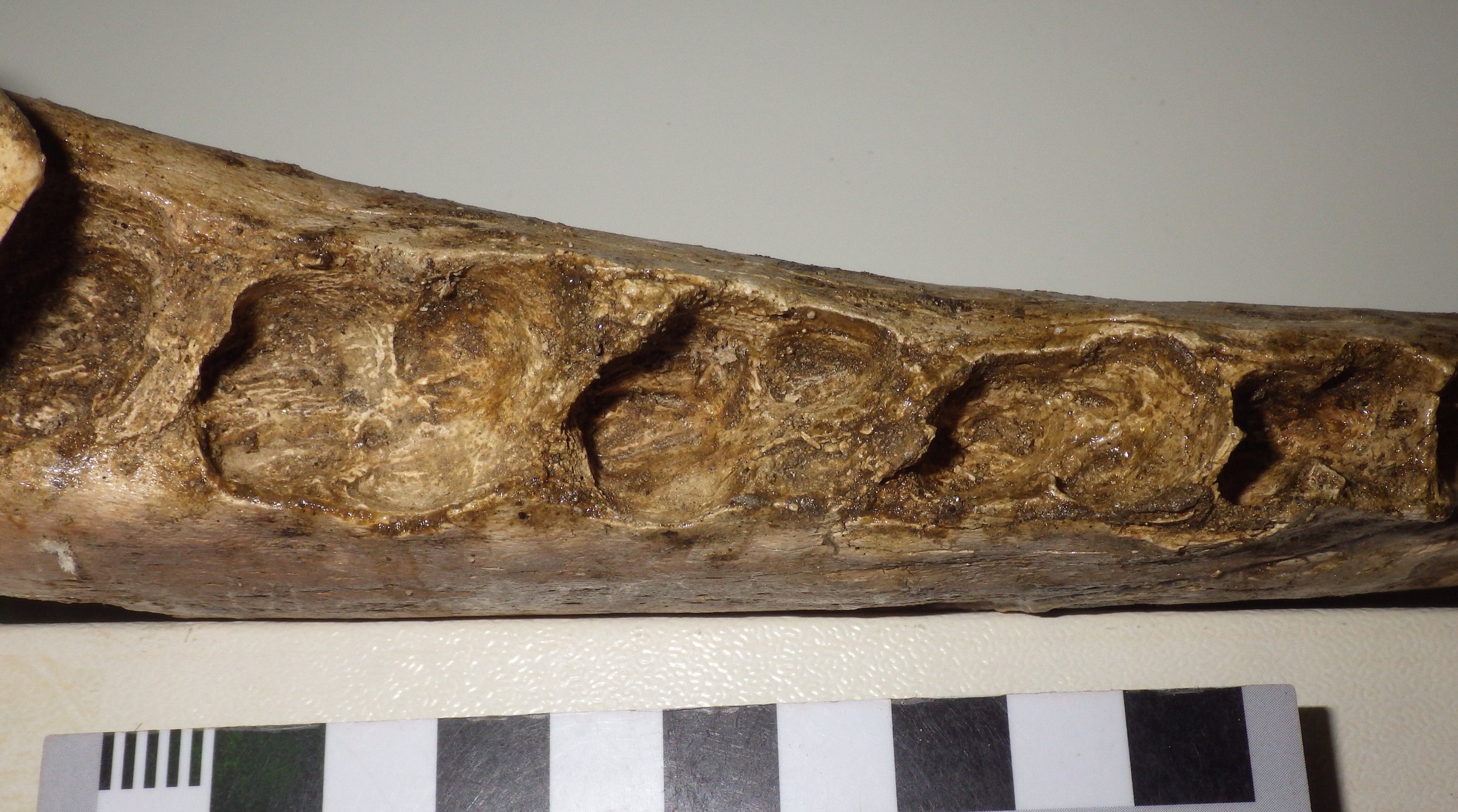
 Even in these views, you may have noticed that the distal end of the bone is a little misshapen. Close-ups reveal that this bone is absolutely riddled with bite marks, presumably from a predator/scavenger gnawing on the bone:


 The broken proximal end also has plenty of bite marks:
 This bone is crying out for a more detailed study. There appear to be at least 2-3 different sets of scratches with different widths. Does this indicate that there were different-sized scavengers? If so, are we looking at different species taking turns (maybe dire wolves followed by coyotes), or different ages of the same species (adult wolves and their pups)? If the scratches show cross-cutting relationships, it might be possible to figure out if the big animals were eating before the small ones, or the other way around, or at the same time.
Fossil Friday - bison jaw fragments
 Some of the species in the Diamond Valley Lake deposits are common enough that it's actually possible to get some idea of intraspecies variability, including growth-based (ontogenetic) differences. Bison are not as common at DVL as horses, but there are still enough specimens to look a bit at age profiles. The specimen above is the anterior end of the lower jaw, with portions of both dentaries. This is a dorsal view, with anterior to the left. Below is a lateral view of the left dentary:
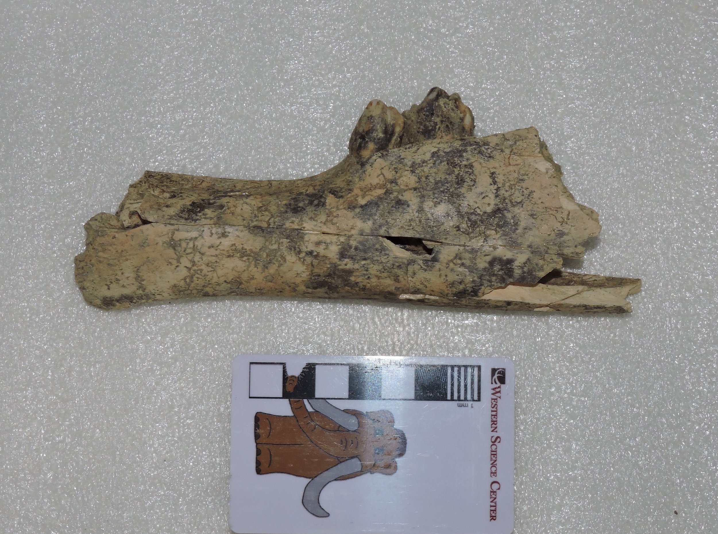 ...and here's right dentary in lateral view:
 Three teeth are preserved, the right 2nd premolar and the left 2nd and 3rd premolars, none of which have any strong indications of wear. The jaw sections are also quite small compared to other bison in our collection. That raises a question: are these teeth permanent premolars, indicating an animal that was maybe 3 or 4 years old, or are they deciduous premolars, in which case it was probably only a few months old? The small size of the jaw actually makes me suspect the latter. I hope at some point to get x-ray images to see if the permanent premolars are present inside the jaw, although this animal could be so young that they hadn't started to develop yet.
Fossil Friday - Bison latifrons skull
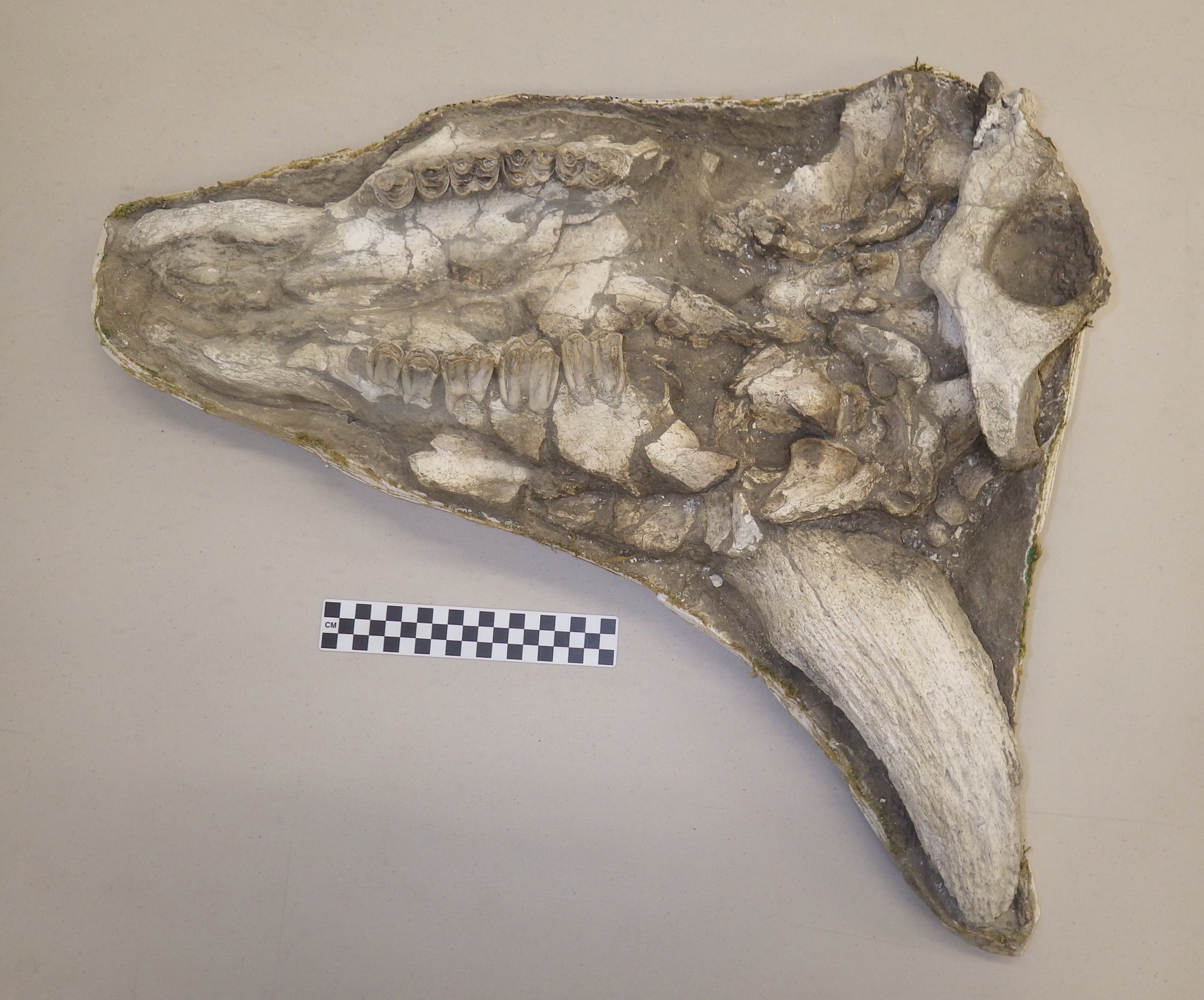
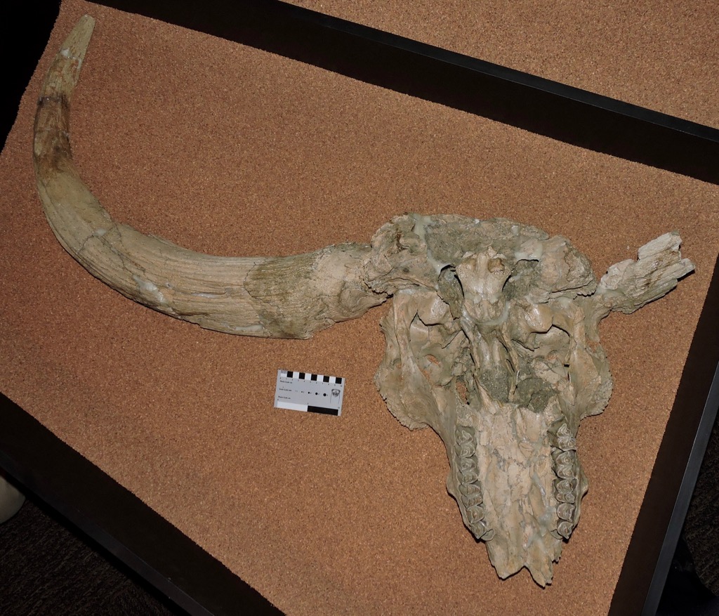 We still have a few days left in our crowdfunding campaign to study mastodons, but for Fossil Friday this week I'm stepping away from mastodons to look at a different animal. Earlier this week the U. S. Government passed a law designating the American bison (Bison bison) as the United States' National Mammal. Bison are prominent in the fossil record, including at Diamond Valley Lake.As I've mentioned in previous posts, there were two species of bison present at Diamond Valley Lake. The more common species was Bison antiquus, a possible direct ancestor to the modern B. bison. The rarer, larger species was the long-horned bison, B. latifrons.Diamond Valley Lake's best B. latifrons skull, shown above, is on permanent exhibit at the museum. It's displayed in ventral view, and in the image the front of the skull is at the bottom. The premaxillae forming the tip of the upper jaws is missing, as is the right horn core, but otherwise the skull is mostly complete if a bit deformed. One thing to keep in mind when looking at fossils of Bison latifrons (and, indeed, any fossil bovid) is that the preserved horn is simply the bone core of the horn. In life the core is covered with a keratin sheath, making the living horn considerably larger.It's somehow fitting that the United States' national mammal is in fact an immigrant to North America, and a relatively recent one at that. Bison were one of the last large mammals from the old world to spread across North America (some of the other large Old World immigrants, such as deer, mammoths, and mastodons, and South American ground sloths, all arrived earlier). In fact, bison only made it south of the 55th parallel (which runs through central Canada) roughly 250,000 years ago, and then rapidly spread across southern Canada and the United States. The first appearance of bison south of the 55th parallel is used by paleontologists at the marker for the start of the Rancholabrean North American Land Mammal Age (NALMA), the last NALMA of the Ice Age.During the Rancholabrean, various bison species ranged across the entire northern hemisphere. While some bison species went extinct at the end of the Ice Age, the genus Bison survived and thrived in the Holocene in both Eurasia and especially in North America. Two extant species are recognized, the American bison (Bison bison) (seen at Yellowstone National Park) and the European bison (Bison bonasus) (seen at the San Diego Zoo Safari Park):
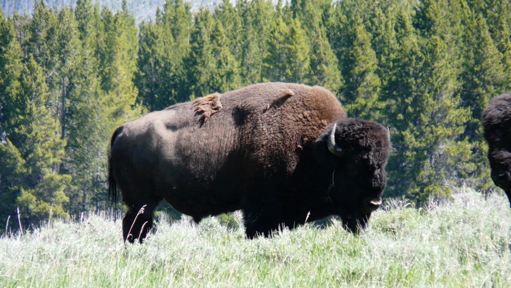
 This wasn't to last, as bison were almost completely wiped out by overhunting and habitat loss over the last few hundred years. Both species have been protected and are slowly recovering, although populations were reduced to such a low number in the past that genetic diversity is quite low. Even so, there are once again wild herds of both species living in protected areas.
Fossil Friday - Bison latifrons horn core
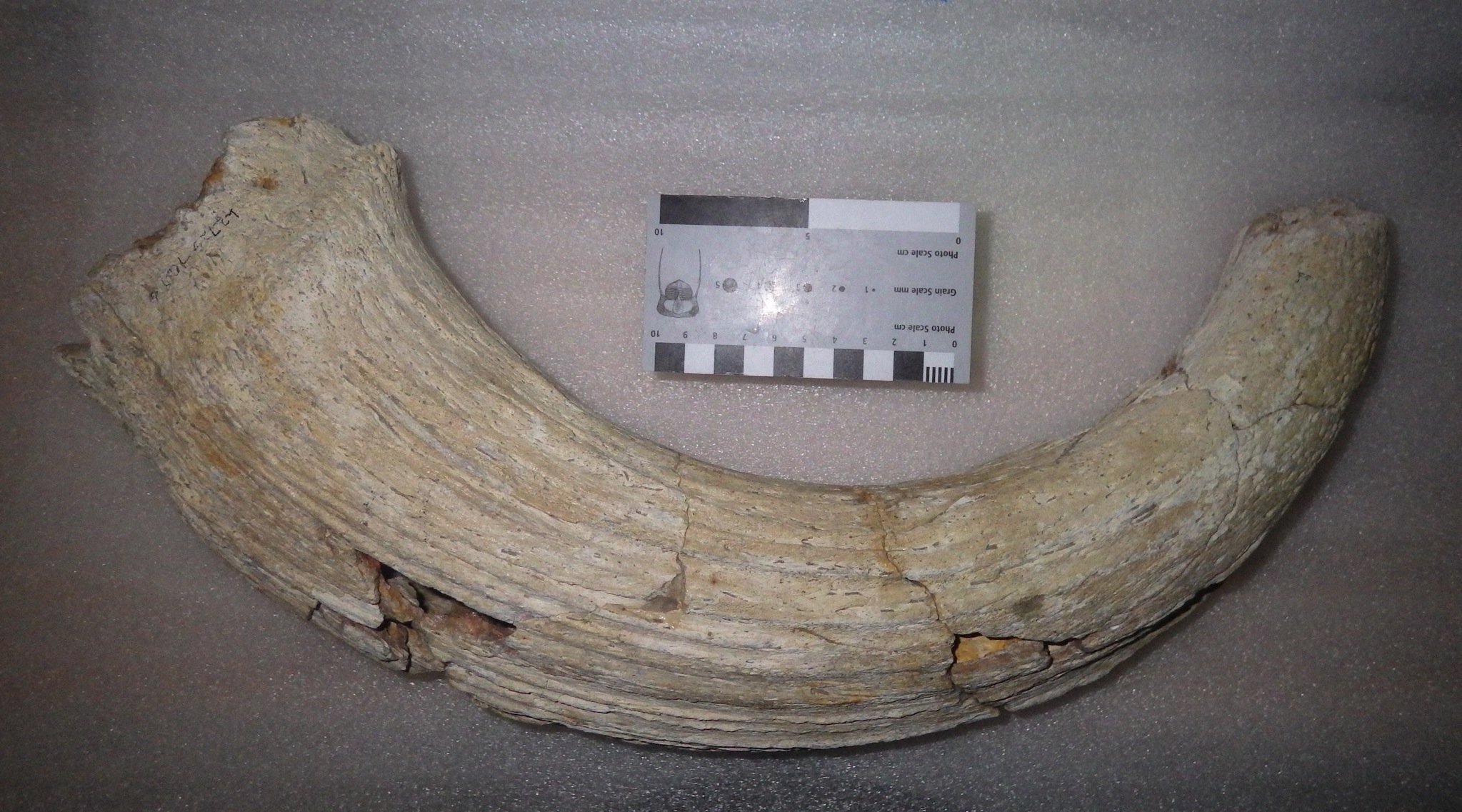
 As I've mentioned in previous posts, there were two different species of Bison present at Diamond Valley Lake, the larger, long-horned Bison latifrons and the (relatively) smaller, shorter-horned Bison antiquus. These species are closely related, and it's often difficult to distinguish between them when dealing with fragmentary remains. But sometimes there's no doubt.Above is the right horn core from Bison latifrons. It has only been partially prepared, and is still in its field jacket. It's shown in dorsal view, with medial to the left and lateral to the right.Like other bovids, bison horns have a bony core that grows from the frontal, one of the bones that makes up the roof of the skull. The lump of bone on the left is part of the frontal. As large as the core is, it's incomplete; at least several centimeters are broken off from the tip. In life, the bony core would have been covered by a keratin sheath, making the horn even larger. And, of course, there was another horn on the left side. All told, it makes for an impressive horn spread, as shown in this cast from the Sam Noble Oklahoma Museum:
Fossil Friday - bison ungal
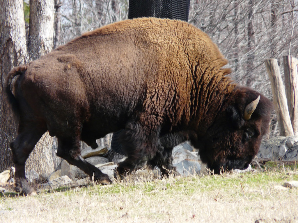
 Hooves are present in several groups of mammals, including the Artiodactyla, the diverse order that includes cattle (bovids) and an enormous range of other animals. Bovids, including bison, have only two toes on each foot, with a hoof at the tip of each toe, as is clear in the photo below of a bison considerately presenting his hooves for inspection:
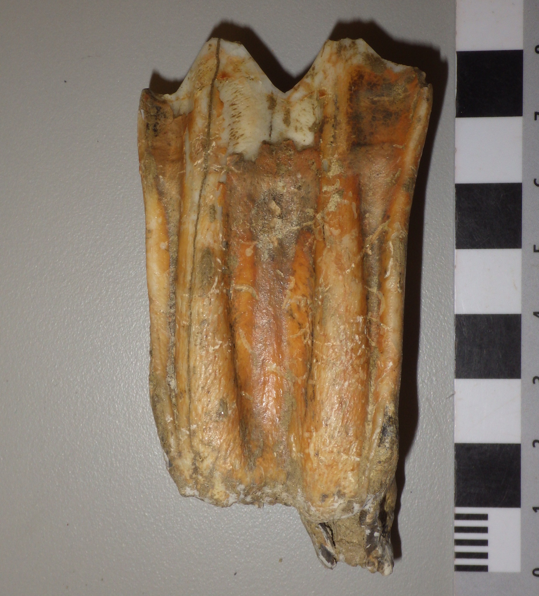
 The outside of the hoof is made largely of keratin proteins, but inside is a bone. This is the last finger or toe bone, known as the terminal phalanx or ungal. The ungal shown above appears to be from left manual digit IV, or the outside toe on the left forefoot. This specimen was collected from the East Dam of Diamond Valley Lake, not far from where the Western Science Center is now located.
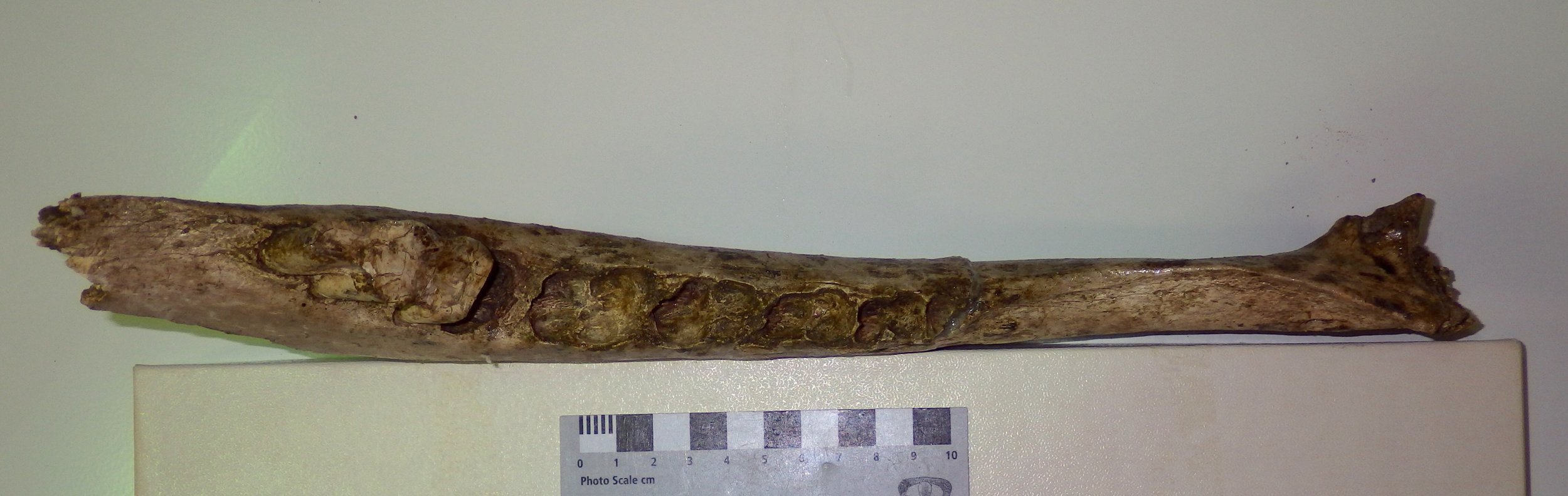
Fossil Friday - bison lower jaw
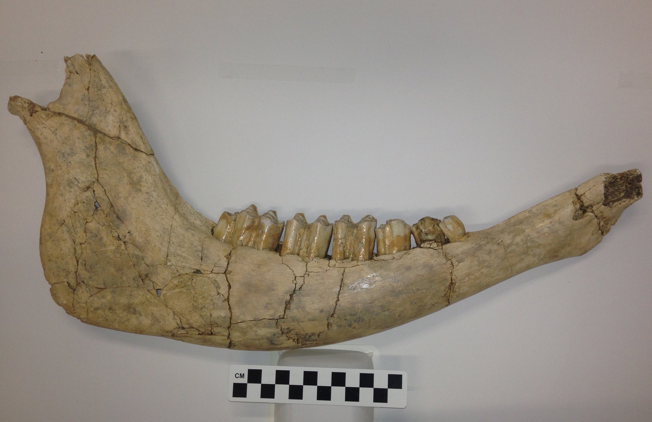 While the Diamond Valley Lake deposits didn't produce anything close to a complete skeleton of a bison, their bones are still among the most common from large animals in the valley, and many of them are well preserved.This is the right side of the lower jaw of Bison antiquus, the smaller of the two Pleistocene bison species from California. Mammals are unique in that each half of the lower jaw is made up of a single bone, called on he dentary, so above is the lateral view of the right dentary. Here's the medial view of the same bone:
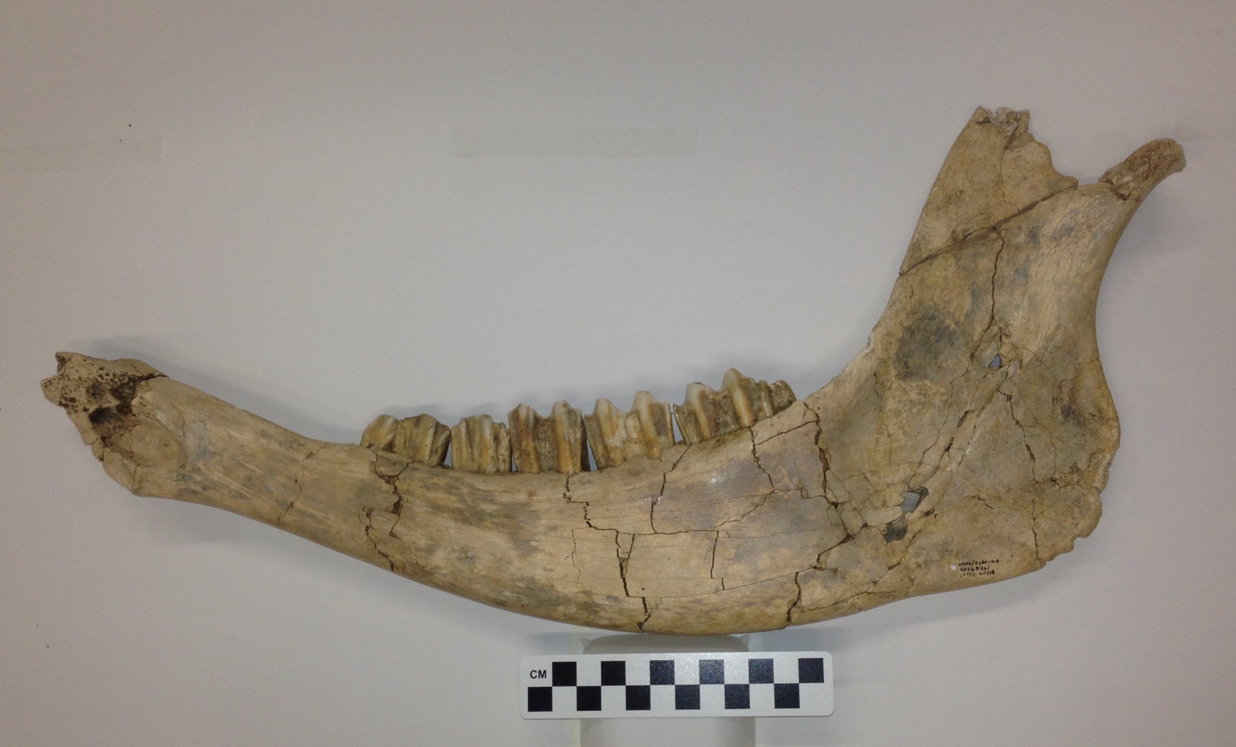 The bone is missing the anterior tip and part of the coronoid process (at the very top), but otherwise is essentially complete. Since the anterior end is absent, the incisor teeth located there are also missing, but otherwise we have the complete dentition. This includes the 2nd, 3rd, and 4th premolars (bison have lost the 1st premolar), and the 1st, 2nd, and 3rd molars. Below is an occlusal view, showing the chewing surfaces of the teeth:
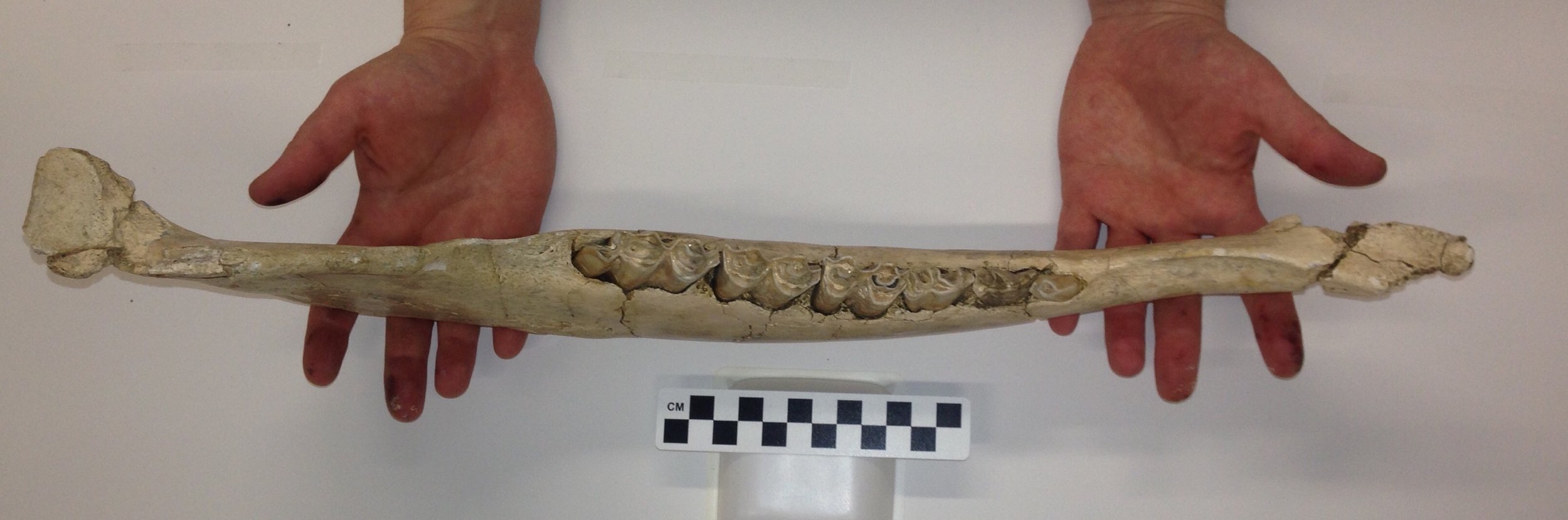 This is a complete set of permanent teeth, so this jaw represents an adult animal, although the moderate wear suggests that it was more likely middle-aged than elderly.This dentary is currently on display at the Western Science Center in the Stories from Bones exhibit.
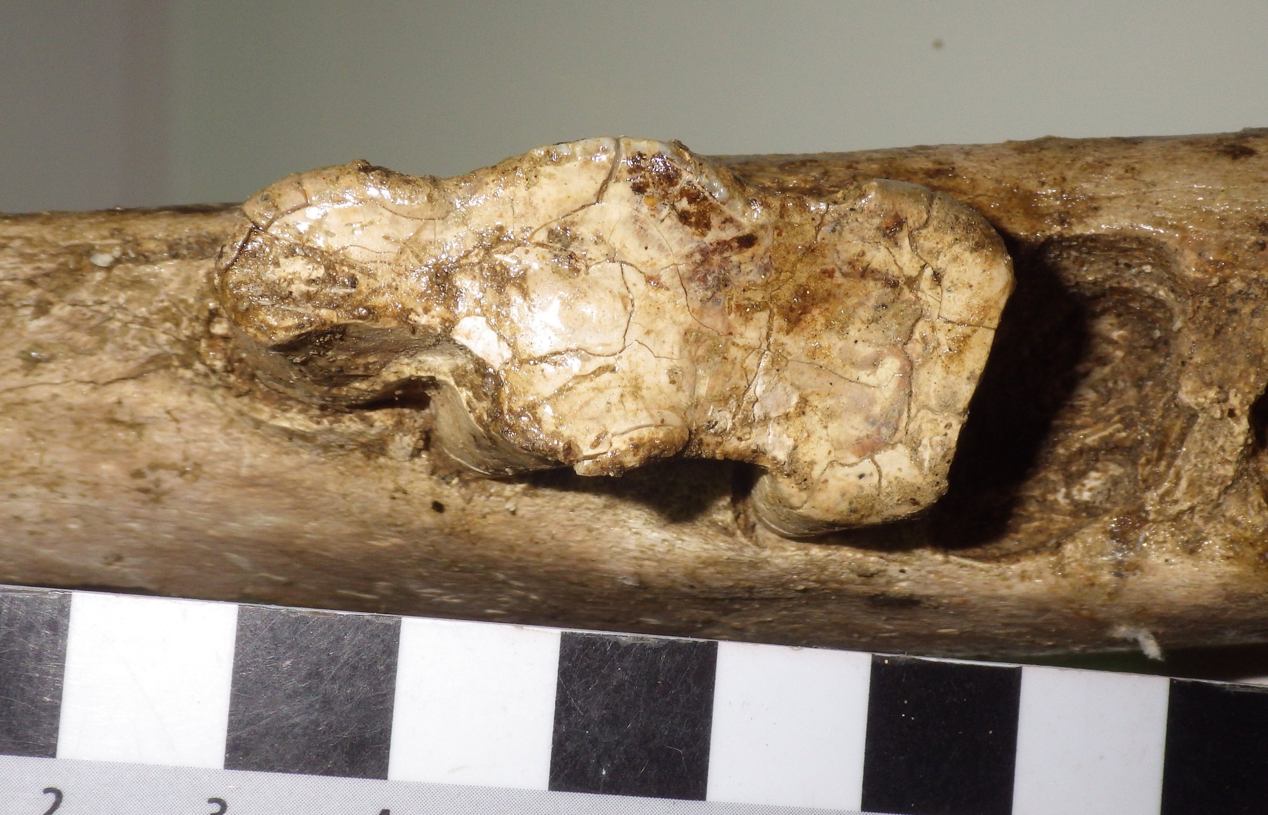
Fossil Friday - bison humerus
 At first glance, bison seem to be rather oddly proportioned, with a relatively massive head and shoulders. And sometimes, things are exactly as they appear; bison really do have huge, heavy heads. Carrying around such a large head has effects on the rest of the body, and bison have strong neck vertebrae, long neural spines on the vertebrae over the shoulders, and robust forelimbs to help support the weight of the skull. This was even more of an issue for Pleistocene bison such as Bison latifrons and B. antiquus, with their relatively larger and heavier horns compared to the modern B. bison.Today's Fossil Friday specimen is part of the right humerus (upper arm bone) from a Pleistocene bison:
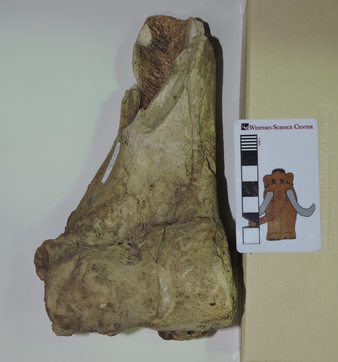 It's shown above from the front (anterior or cranial view). This is only the distal part of the bone, with the elbow joint at the bottom. The proximal half with the ball joint at the shoulder is missing. This is a big, heavy bone; the articulation surface at the elbow is about 9 cm across.Here's the same bone from behind (posterior or caudal view):
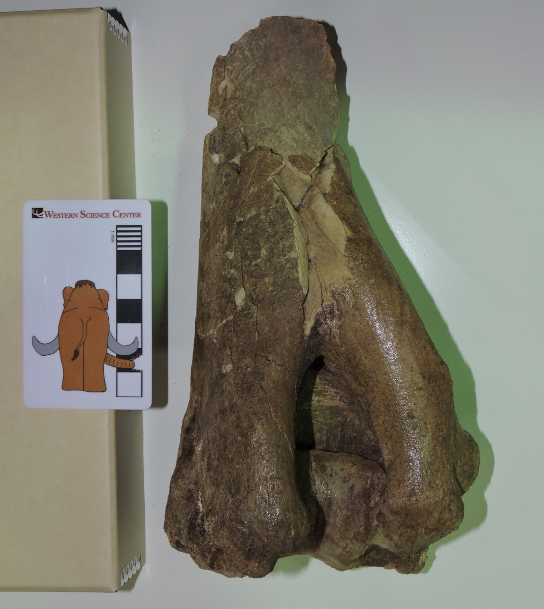 The large notch at the bottom is called the olecranon fossa. It accommodates a large projection on the connecting bone, the ulna, called the olecranon process. This process forms the point of the elbow, and serves as the attachment point for the triceps brachii muscle that extends the forelimb. When the triceps brachii is flexed, it straightens the forelimb, rotating ulna's olecranon process into the humerus' olecranon fossa.
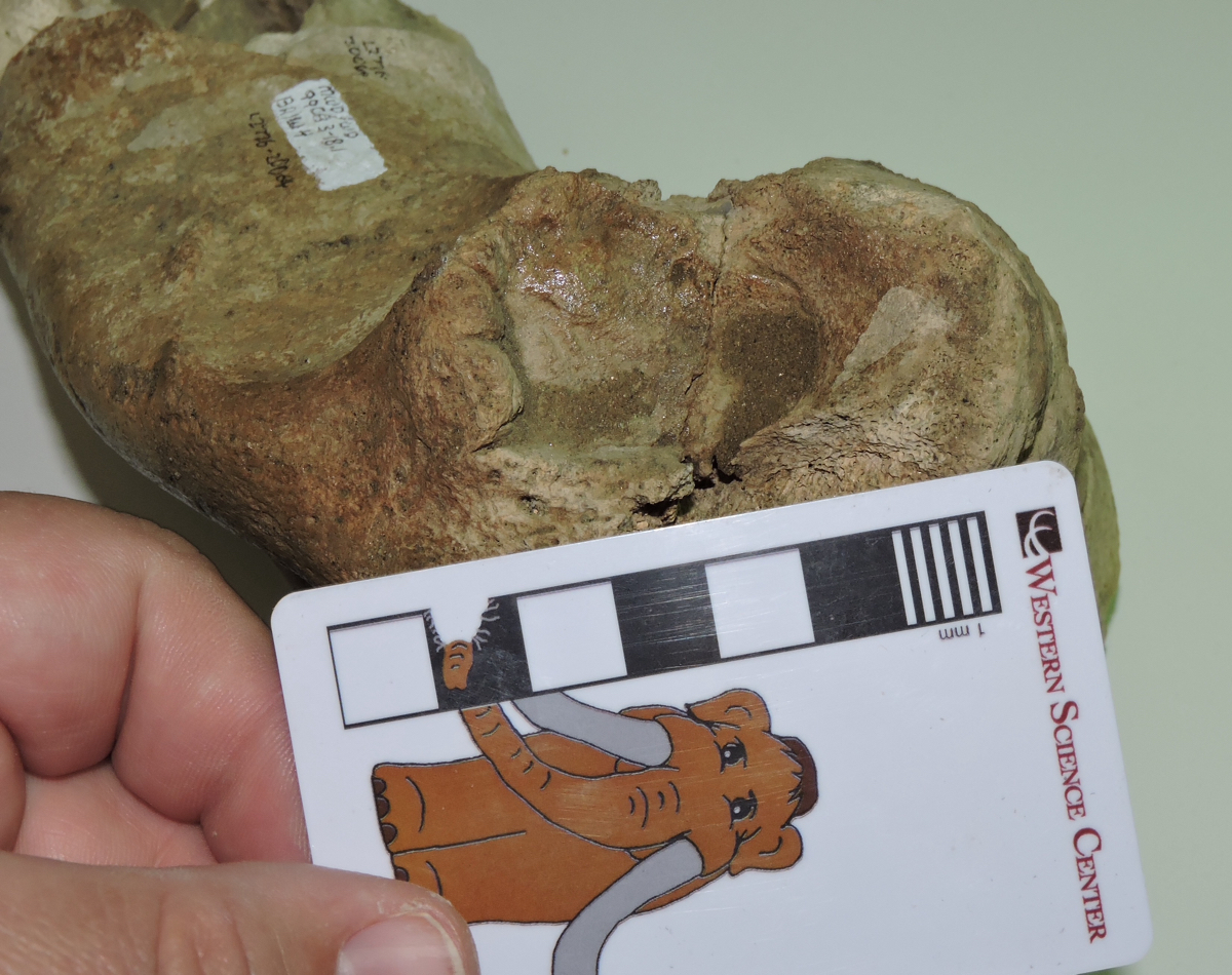 Above is an oblique view of the lateral side of the humerus. The circular area above the scale bar is called the lateral epicondyle, and in this specimen it has a somewhat irregular shape and texture. This could be due to osteoarthritis, which wouldn't be surprising in the elbow joint of a heavy-headed animal such as a bison. The distal end of this humerus is fully fused to the rest of the bone (unlike the mastodon humerus we featured last week), suggesting that this bison was full grown at the time of its death.
Fossil Friday - Stories from Bones exhibit
![]() For Fossil Friday this week, I want to highlight Western Science Center's new exhibit "Stories from Bones", which opens tomorrow.While WSC has excellent paleontology exhibits, as with any museum with a large collection many of the specimens are not on public display. There are a variety of reasons for this. Of course, the biggest obstacle is money; cases, information panels, interactive, floor space, and other requirements for an effective display are all expensive, and even the healthiest museums operate on a shoestring budget. Besides money issues, many specimens are just not suitable for display. Perhaps they're too fragile to risk moving around too much, or too fragmentary to interpret for the public (a specimen that visually looks like a piece of junk can still produce valuable scientific data). Even with all these limitations, we strive to make as much of our collections accessible to the public as possible. "Stories from Bones" is a result of that effort.
For Fossil Friday this week, I want to highlight Western Science Center's new exhibit "Stories from Bones", which opens tomorrow.While WSC has excellent paleontology exhibits, as with any museum with a large collection many of the specimens are not on public display. There are a variety of reasons for this. Of course, the biggest obstacle is money; cases, information panels, interactive, floor space, and other requirements for an effective display are all expensive, and even the healthiest museums operate on a shoestring budget. Besides money issues, many specimens are just not suitable for display. Perhaps they're too fragile to risk moving around too much, or too fragmentary to interpret for the public (a specimen that visually looks like a piece of junk can still produce valuable scientific data). Even with all these limitations, we strive to make as much of our collections accessible to the public as possible. "Stories from Bones" is a result of that effort.
 Mammoth jaw display in "Stories from Bones".
An important aspect of planning an effective exhibit is developing a theme. An exhibit is telling a story, and you need to be aware of what that story is as the exhibit is being designed. The theme might be "We have a bunch of stuff!", but while that was a common theme in museums a century ago (and one I personally appreciate), it does not generally make for the most informative exhibit experience for the majority of visitors.Once the theme is established, it's important to stick to it, so that the exhibit story remains coherent. Imagine reading a mystery novel in which three chapters are devoted to a history of the development of the gunpowder used in the crime, and an additional chapter describes the etymology of the last name of the victim, when neither is important to the outcome of the story. Each of these things might be individually interesting, but if you try to talk about all of them then you risk obscuring everything. There is a real risk of this "mission creep" in an exhibit based on a data-rich field such as paleontology. We might talk about evolutionary relationships, paleoenvironmental indicators, biogeographic information, site-specific descriptions, or an array of other things. Talking about any of these might be a good idea; talking about all of them is a bad idea.The permanent paleontology exhibit at WSC does this very well. The exhibit is basically a review of the Diamond Valley Lake Local Fauna; what was here, how does it compare to the rest of Southern California, and (as a secondary point) what does it tell us about the local Pleistocene paleoenvironment. In contrast, "Stories from Bones" asks "What do these fossils tell us about the lives and deaths of these individual animals?".To that end, "Stories" has a series of displays that talk about how paleontologists determine how old an animal was when it died. We have several cases that look at tooth replacement in proboscideans, horses, and bison, such as the two mammoth jaws above (they're close to the same size, but one animal was about 30 years older than the other), or the three bison dentaries shown below that represent young, middle-aged, and elderly animals.
 Bison jaw display in "Stories from Bones".
We have several examples of bones that were broken and healed, evidence of events that took place during an animal's life:
 Broken and healed bones in "Stories from Bones".
We also have several cases that describe taphonomic features, looking at what happened to an animal at or immediately after death.We designed and built a number of interactive displays for this exhibit. The most prominent is a cast and video of the CT scans of Max the Mastodon's lower jaw, taken back in August.
 Max's CT-scan station in "Stories from Bones" during installation, under the watchful eye of @MaxMastodon.
We're proud of the fact that several of our interactive displays ask visitors to map or measure specimens and reach conclusions based on their data:
 A more extensive version of the bison tooth display shown here is also available as a guided activity for school groups visiting the museum, and as a kit available for purchase.If you're a regular reader of this blog, you'll find that "Stories from Bones" draws heavily from my past "Fossil Friday" posts. For most of those specimens, this is the first time they've ever been on public display, so if you're near Southern California make sure to stop by the museum. "Stories from Bones" opens on October 31, and will remain open into May 2016.
Fossil Friday - bison metacarpal
 This week's Fossil Friday features a metacarpal (hand bone) from a Pleistocene bison. The example shown here is a left metacarpal shown in anterior view, with the proximal end (at the wrist) on the left and the distal end (at the first knuckles) on the right.Like many other advanced artiodactyls, in bison this structure is actually two bones that are fused together. These are the third and fourth metacarpals, the hand bones that in humans support the middle finger (Digit III) and the ring finger (Digit IV). Both of these "fingers" are present in bison as well, with each supporting a hoof (remember that our hands are just highly modified front feet). The articulations for each digit are visible at the distal end of the bone, on the right side of the picture.Here's the same bone in posterior view (the equivalent of "palmar view" in a human hand):
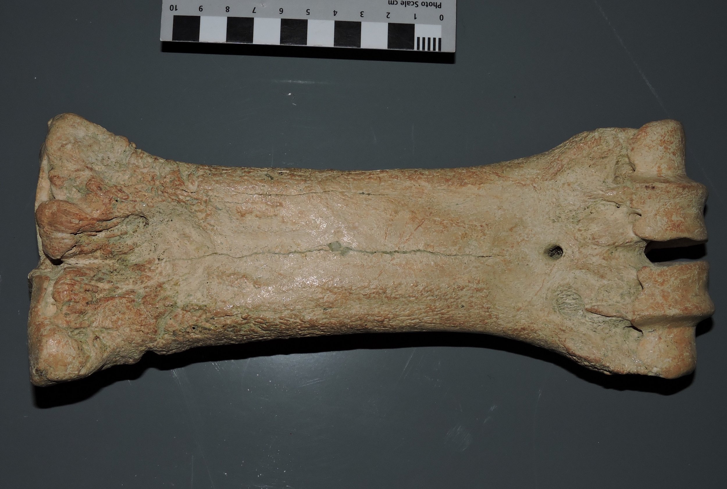 In this view the articulations for digits III and IV are more clearly visible on the right, but the groove indicating the line of fusion between the third and fourth metacarpals is almost completely lost.Last year I posted photos of the metacarpal from the extinct camel Camelops. Camels and bison are not very closely related to each other, but they are both artiodactyls that show similarities is the hand and foot structure. In both animals the front foot is reduced to just the fused third and fourth metacarpals. Camelops was a large animal that was much taller than Bison, and this is reflected by the relatively long, slender metacarpal. Bison, while shorter, were heavier and had to support a massive head, resulting in a shorter, stouter metacarpal.
Fossil Friday - Bison atlas vertebra
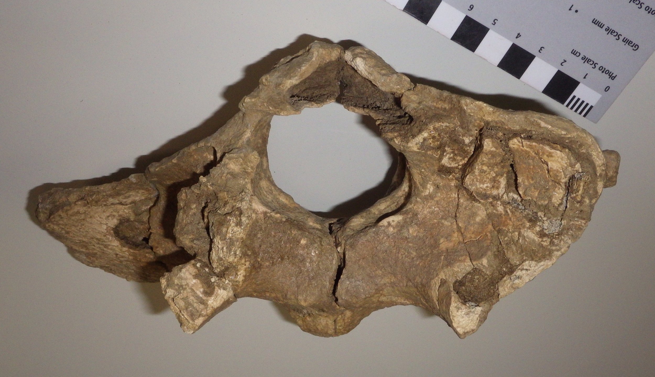 For this week's Fossil Friday I'm going to stick with bison, this time featuring an atlas vertebra.As I've described in earlier posts, the mammalian vertebral column is generally divided into five different regions. The most anterior are the cervical (neck) vertebrae. Almost all mammals have exactly seven cervical vertebrae, but mammals are unique among vertebrate classes in having such a tightly constrained cervical count. The first two cervical vertebrae, the atlas and the axis, are always highly specialized and look very different from any of the other vertebrae.That is mainly because of the complex articulations necessary to allow a range of movement in the head. When you nod your head (flexion and extension), that motion primarily occurs at the joint between the front of the atlas vertebra and the skull. When you shake your head (rotation) the motion is mostly at the joint between the back of the atlas vertebra and the front of the axis vertebra. That all means that the atlas has to accommodate one type of motion (nodding) on its front surface, a different motion (rotating) on its back surface, while providing attachment points for all the muscles that actually cause the movement, and allowing a passageway for the spinal cord and the vertebral arteries and veins through the middle of all of this movement. It's no wonder that the atlas has such a distinctive appearance. (As an interesting aside, whales, which have almost no ability to move their heads, still have complex and unique atlas and axis vertebrae, a relict of their ancestors that had much more mobile necks.)So, with all that said, the image at top shows a bison atlas vertebra in anterior (cranial) view. The bone is somewhat crushed and incomplete; much of the right side in particular is missing (remember, since this is anterior view, the right side of the vertebra is on the left side of the photo). The large hole in the bone is the neural canal, the passageway for the spinal cord. Below and beside the neural canal (centered at the "5-o'clock" and "7-o'clock" positions) are large depressions that articulate with the occipital condyles on the skull; this is the main "nodding" articulation.
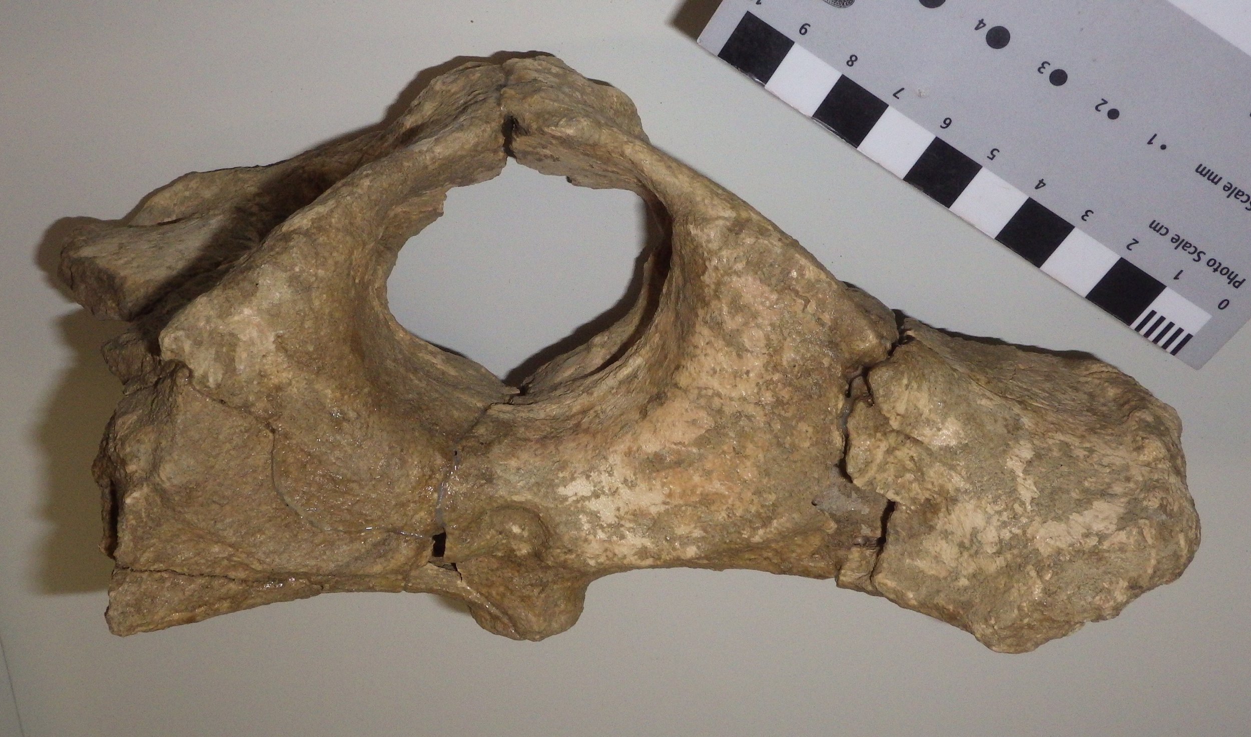 Looking at the same vertebra in posterior (caudal) view, we see broad, flat surfaces below and beside the neural canal. These are the articular surfaces for the axis vertebra, where rotation occurs. The large projection of bone on the right is a lateral or transverse process (the corresponding one on the left side is broken off). This provides a surface for the attachment of neck muscles associated with head movement. Bison have large, heavy heads, and so the muscle attachment areas to support them are relatively large.Here's the atlas in dorsal (top) view:
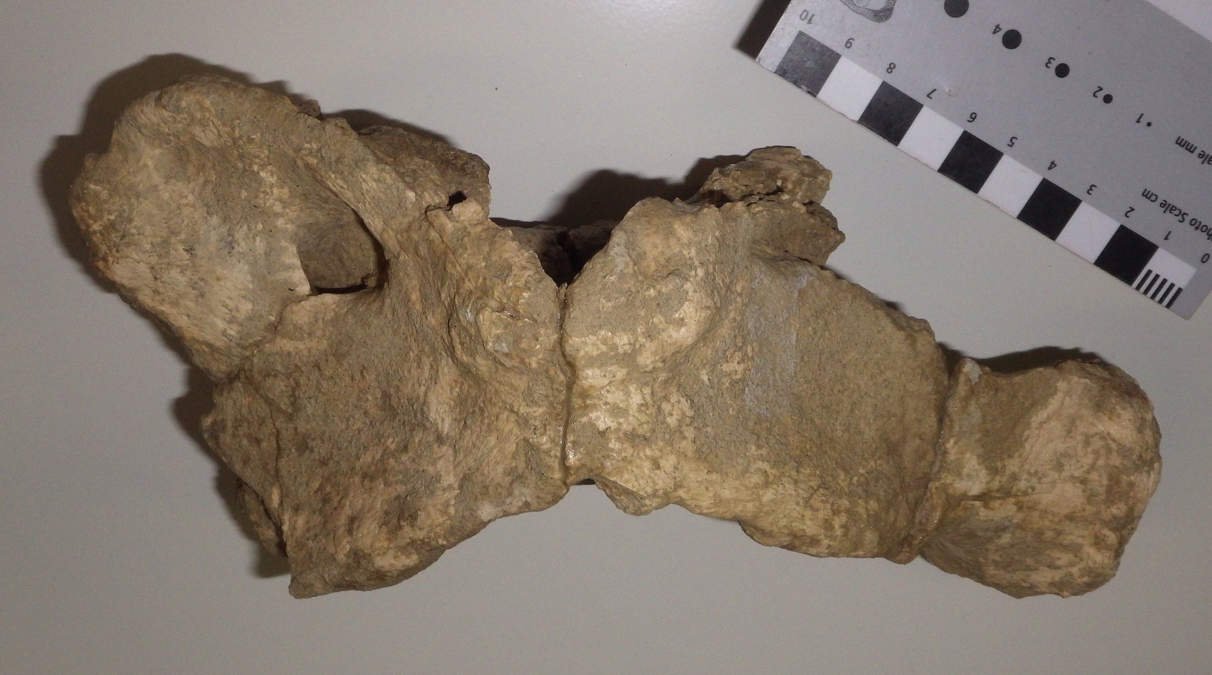 At this angle you can see that the transverse process is broad across the top surface, and has quite a large surface area. The opening toward the upper left is the alar foramen (the right one is only partially preserved), which provides a passageway for the vertebral arteries and veins.Even allowing for damage to the bone, the shape of this atlas seems a bit different from that of the modern Bison bison (see this site for an example). There are two different species of Bison known from the Diamond Valley Lake fauna, B. antiquus and B. latifrons. B. latifrons has a larger, heavier head than B. antiquus, and it's possible that this might be reflected in the anatomy of the cervical vertebrae, but I've not yet checked references to confirm this. At the moment, we have this particular atlas identified as Bison sp.
Fossil Friday - "Then the rats got him"
 Bison are among the most common large animals in the Pleistocene Diamond Valley Lake fauna, but like almost all the remains from these deposits they are usually fragmentary. But even fragmentary fossils can provide a lot of information, including the bison right lower jaw fragment shown here.Sometimes bonebeds or other rich deposits of fossils are formed during catastrophic events, with large numbers of organisms buried together very rapidly. That's not what we see in Diamond and Domenigoni Valleys, however; these deposits are primarily the result of the gradual accumulation of skeletons over thousands of years. That means that an individual skeleton may have laid exposed at the surface for a significant period of time before it was eventually buried. This exposes the bones to sun, wind, and rain, all of which can cause the bone to deteriorate and the skeleton to fall apart, and to scavengers, which may damage or eat the bones, or carry pieces away. All these things contribute to the fragmentary nature of the Diamond Valley Lake specimens.Evidence of the specific types postmortem trauma experienced by these bones is sometimes preserved. If we zoom in on the bison jaw shown above, we can see a series of grooves cut into the bone:
 These grooves are bite marks, most likely made by rodents. Bone gnawing has been documented in lots of different rodents, including rats, mice, and squirrels. It's generally attributed to the need for rodents to keep their ever-growing incisors from becoming too long, and perhaps to provide an additional source of calcium. But the presence of these marks immediately tells us quite a bit of additional information; the bison died in a setting that didn't result in immediate burial, and rodents were present in the environment.Of course, from the bison's point of view, maybe this is only adding insult to injury!
Fossil Friday - bison lower jaw
 I'm in Chicago this week for the National Science Teachers' Association meeting, in part so that my wife Brett and I can demonstrate teaching kits that we're developing based on the WSC collections (I'll have more about those in a future post). One of these teaching kits involves fossil bison, including the specimen featured in today's Fossil Friday. This is a partial right lower jaw from a bison, seen above in lateral (side) view. Below is the same specimen from above (dorsal view):
 While most of this jaw is missing, it does preserve the tooth sockets ("alveolae"), even though only a single tooth is preserved. That tooth is the last molar, m3, and it's pretty unusual in occlusal view:
 The tooth is almost completely worn away. None of the usual complex enamel patterns that we see in bison teeth are present, because the enamel is mostly gone, except for a few patches on the edges of the tooth. Modern bison live to about 15 years in the wild, and this one had to be getting up there.But there's actually more to look at here. It's not that unusual to have a jaw fragment with only one tooth preserved; once a jaw is broken it's easy for the teeth to fall out. But if we look at the empty tooth sockets in this jaw, we can see that's not what happened here:
 These sockets actually have bone covering the openings. That means that the teeth fell out before the animal died, and the sockets healed over! This bison was shockingly old; it had actually lived long enough for every tooth except the last one wear all the way down to the roots and fall out, with the sockets healing over. Even the last tooth was close to falling out when the animal died.It's tough to estimate the age of such an old animal, but this had to be a particularly elderly individual, and has resulted in us nicknaming it "Methuselah".
Fossil Friday - Bison antiquus skull
 Bison are among the most common large animals from the Diamond Valley Lake region, and we have a number of nice specimens at the Western Science Center. Shown above is a cranium of Bison antiquus, one of several such skulls in our collection.This skull, while incomplete, is in pretty good shape. It's still in its field jacket, and only partially prepared, so only the ventral (bottom) side is visible. The front of the skull is to the left. The left horn core is noticeably missing, and there is some crushing of the skull that's most clearly visible along the right side (bottom of the image). The 2nd premolar is missing on each side, but the other teeth are all present. If you're looking carefully, you might have noticed that there are no incisor teeth in the front of the mouth. Bison don't have incisors in the upper jaws, a character they share with other "horned ruminants" (Infraorder Pecora). The mass of bone at the back of the skull (on the right side of the image) is the atlas (first neck vertebra), more-or-less still attached to the skull.There are two different species of bison present in the Pleistocene deposits at Diamond Valley Lake, the smaller Bison antiquus and the larger, longer-horned Bison latifrons. (When I say "smaller", that's relative; B. antiquus was still larger than modern bison.) Bison antiquus seems to be the more common of the two fossil species in this area. It's generally thought that B. antiquus is the direct ancestor to the modern bison, Bison bison.
Fossil Friday - bison horn
 Bison are among the most common animals in the Diamond Valley Lake Local Fauna. While no complete bison skeletons were recovered during excavations for the construction of the reservoir, large numbers of fragments, individual bones and teeth, and associated bones were discovered. Some of these are impressively large, including the horn core shown here.Bison are members of the Family Bovidae, a diverse group that includes cattle, goats, sheep, and antelope. A characteristic shared by practically all bovids is the presence of horns in males (there are some domestic bovid breeds in which the males lack horns); in many species the females have horns as well. The horns vary greatly in size and shape between species, but they always have certain features in common: they form as outgrowths of the frontal bones on the roof of the skull, they do not branch, and they are covered in a keratin sheath. Keratin doesn't usually preserve in fossils, so what we generally see in fossils is the rough bone underneath. Because the horn is part of the frontal bone, it is not shed each year like the antlers of deer.There are two different extinct species of bison known from Diamond Valley, Bison antiquus and Bison latifrons; among other differences, B. latifrons is larger and has much longer horns. As big as this specimen is, it's not immediately clear which species it represents. It's on the small side for B. latifrons, but a little larger than some of our B. antiquus specimens.
Fossil Friday - bison molar
 As our molding and casting program gets underway, we've been going through the WSC collections looking for good candidates for molding. We've been particularly interested in teeth, because in many mammals the teeth are highly distinctive and potentially provide a lot of information about the animal. Over the last week we've been focusing on bison teeth.Shown above is a more-or-less complete lower right molar from Bison, probably the 2nd molar, shown in labial view. Here's the same tooth in lingual view:
 In this view there's a ridge running vertically down the middle of the tooth called a stylid. This is a column of enamel that will show up on the occlusal surface of the tooth as a small ring of enamel if the tooth is sufficiently worn. This stylid seems to always be present in Bison, but in the closely related genus Bos (cows) the stylid is usually absent or only weakly developed.Here's the tooth in occlusal view:
 Bison have a selenodont tooth pattern with two main crescent-shaped cusps, similar to many other artiodactyls. The tooth is worn, but only slightly, so this animal was likely a young adult when it died. The tooth had not yet worn down as far as the stylid on the lingual surface, so the enamel loop isn't visible; had the animal lived longer the loop would eventually have appeared as the tooth wore down.This tooth was found near the east dam of Diamond Valley Lake.
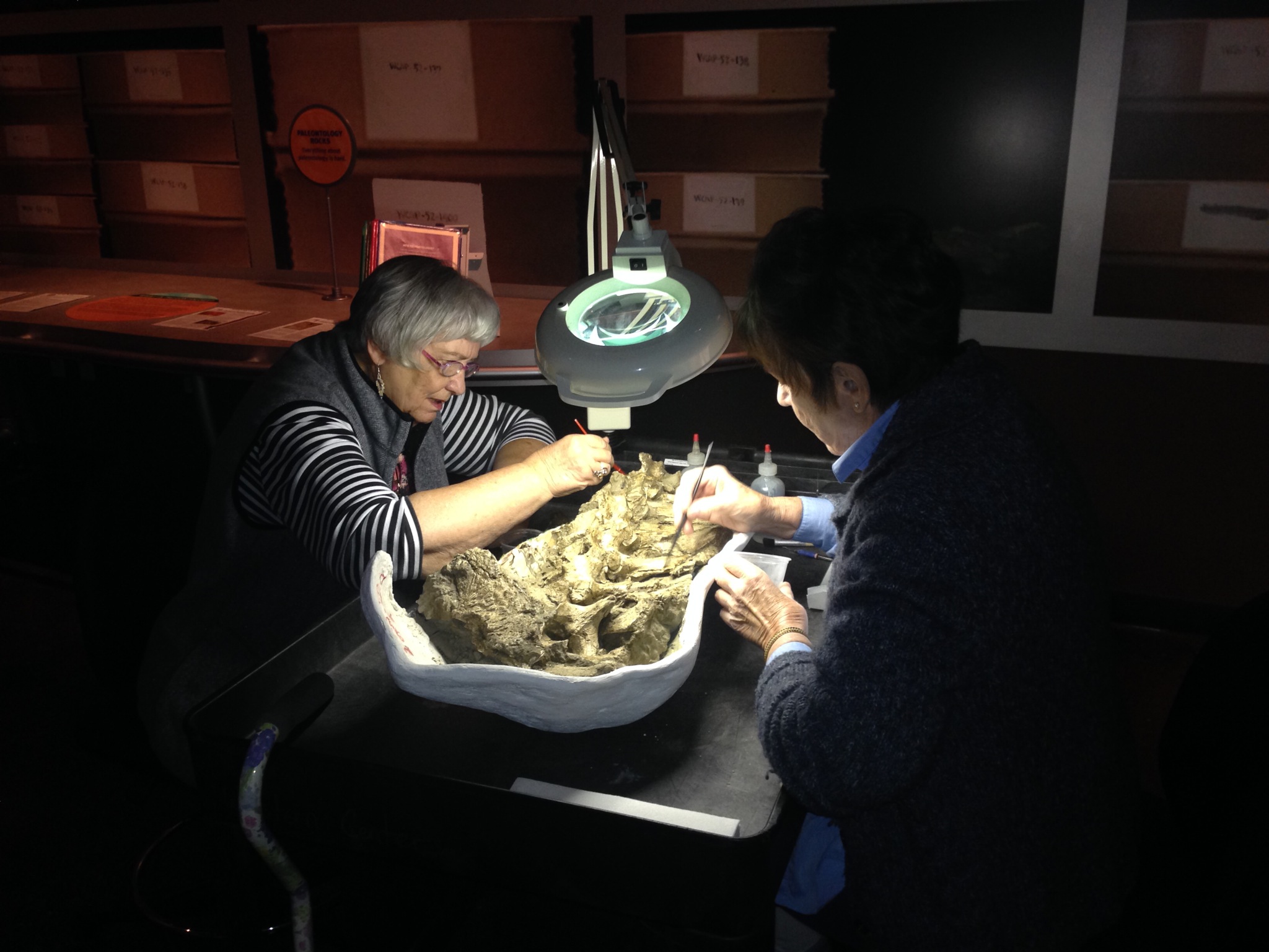
Bison vertebrae on the prep table
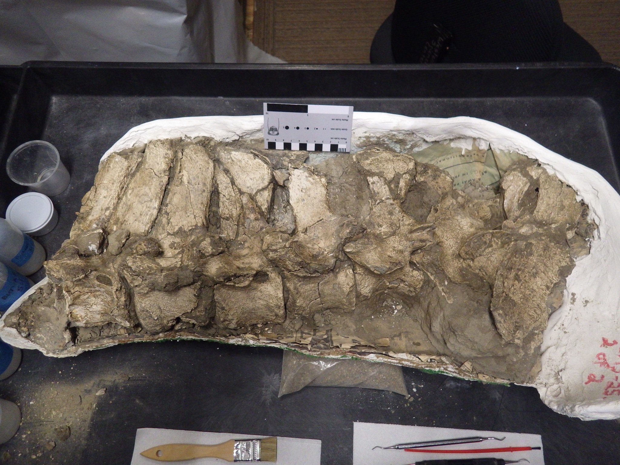 Last week we started a new preparation project, a roll-out cart where we do fossil prep work on the exhibit floor. Our first project for the cart is a jacket containing a series of bison vertebrae.The way the jacket has been opened, the vertebrae are exposed on the left side, so they're seen above in left-lateral view, with anterior to the left. As I mentioned a couple of weeks ago, mammal backbones are generally divisible into five different regions based on their position and particular features.This small sequence actually contains vertebrae from three of the five different regions, as shown in the annotated image below. Bones outlined in red are thoracic vertebrae, those in blue are lumbars, and the ones in green are sacrals and the associated hip bones:
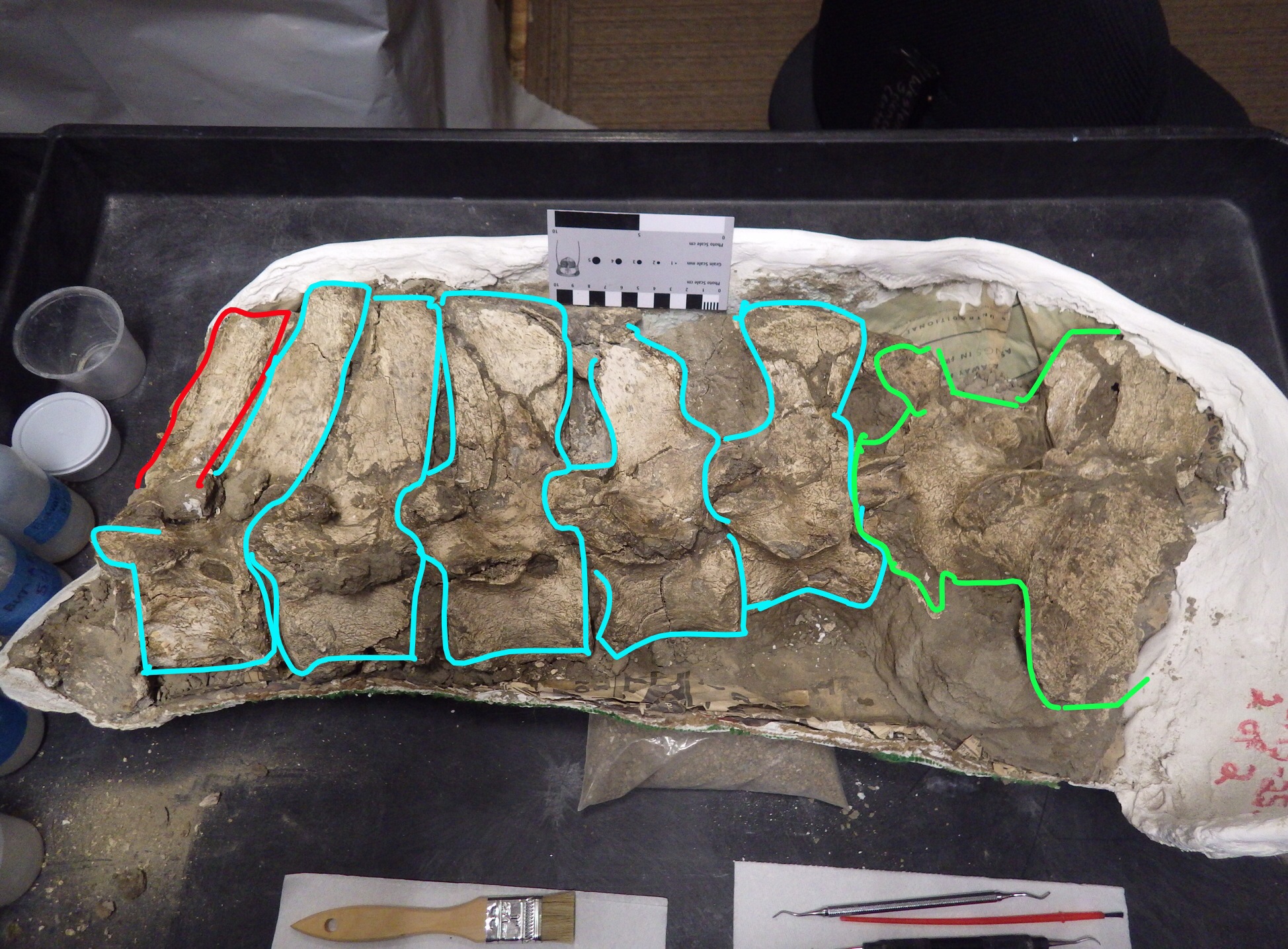 Typically, bison have 14 thoracic vertebrae. We have part of the last one, the extra neural spine at the front of the jacket. We also have part of the main body (the centrum) of this vertebrae, but it's separated from the jacket.Bison generally have five lumbar vertebrae, so we have the entire lumbar series preserved (compare them to this image of a modern bison lumbar series, shown in dorsal instead of lateral view). We are missing the long transverse processes that should be sticking out of the left side of each vertebrae; it's not unusual for them to break off before burial.Bison also have five sacral vertebrae that are generally fused to each other and to the hip bones (at least in adults). We have at least the first sacral vertebra, and some additional portion of the sacrum, but that area is still kind of a mess and it will be awhile before we know exactly what's in there.I'll be posting periodic updates on our progress with this jacket as preparation continues. The preparation cart is currently on display at Western Science Center from 10:00-12:00 on Tuesdays, with additional times to be added soon.
WSC's Fossil Preparation Exhibit
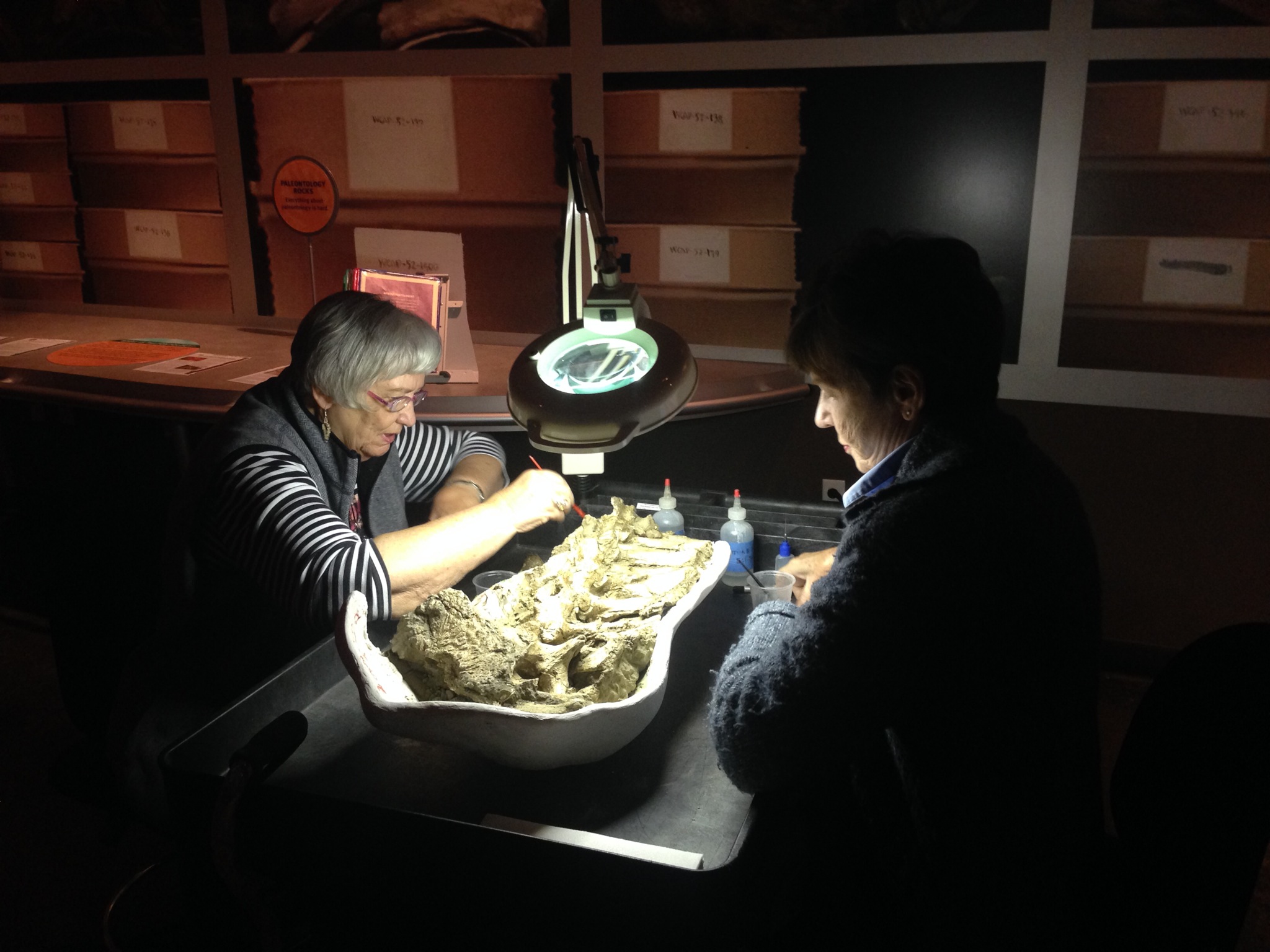 Like most collections-based museums, the Western Science Center has far more specimens than we could ever put on exhibit. We want to make our collections and procedures accessible to as many people as we can, but there are all kinds of technical, financial, and security hurdles. Even so, we're always exploring new ways to accomplish this, and today we're launching our latest effort — a fossil preparation demonstration area on the exhibit floor.Many natural history museums now have "fish-bowl"-style preparation labs so that visitors can observe staff and volunteers cleaning specimens. We hope to open such a lab one day at WSC, but that will take a lot of time and money and I'm not willing to wait. So we've put together a rolling preparation cart that our lab volunteers can take to the exhibit floor to do their work while answering questions and giving visitors an up-close look at the fossils.Two of our lab volunteers, Phyllis and Margit, have agreed to help us test out this new system by doing their prep work on the exhibit floor. Their initial specimen is an articulated series of seven Bison vertebrae that were partially prepared but are still in their field jacket.
 We're going to be testing and refining this system over the next few months. The preparation cart will be on the exhibit floor whenever we have staff or volunteers available to do preparation work, initially on Tuesdays from 10:00-12:00 but with additional hours to be added later.