
 This may look a bit like a zombie hand reaching from the grave in some B-horror movie (at least it does to me). In fact, it's a fossil that's quite a bit more interesting and less dangerous than a zombie hand.This is part of the lower jaw of the western camel Camelops hesternus, a fairly common species across western North America in the Late Pleistocene. This is the chin, technically called the mandibular symphysis, seen from underneath (ventral view). The forked structure on the left is behind the symphysis, where the left and right dentary bones separate to form each side of the lower jaw. The "fingers" on the right are the incisor teeth.Here's the dorsal view:
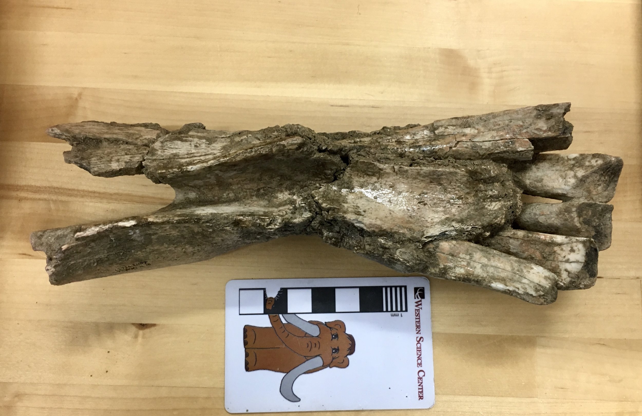 The incisors are more clearly visible in this view. Camels have three lower incisors on each side of the lower jaw; this specimen preserves all three on the right side, but only the first two on the left.Below is a lateral view of the right side:
 Because the bone is damaged on this side the entire length of the 3rd incisor is visible (minus whatever part of the tooth wore away before the camel died). Camels eat a tough, abrasive diet that is hard on teeth; camels compensate for this in part by having really long incisors that take a long time to wear down (the technical term - because OF COURSE there's a technical term! - is hypsodonty).We have more of this jaw, and it appears that there are no lower canine teeth. Since this is an adult animal, I suspect that it represents a female, since male camels (including Camelops) tend to have fairly prominent upper and lower canines. However, I need to do a little more research on Camelops to confirm that it displays this type of sexual dimorphism.Here's a close-up of the occlusal surfaces of the teeth:
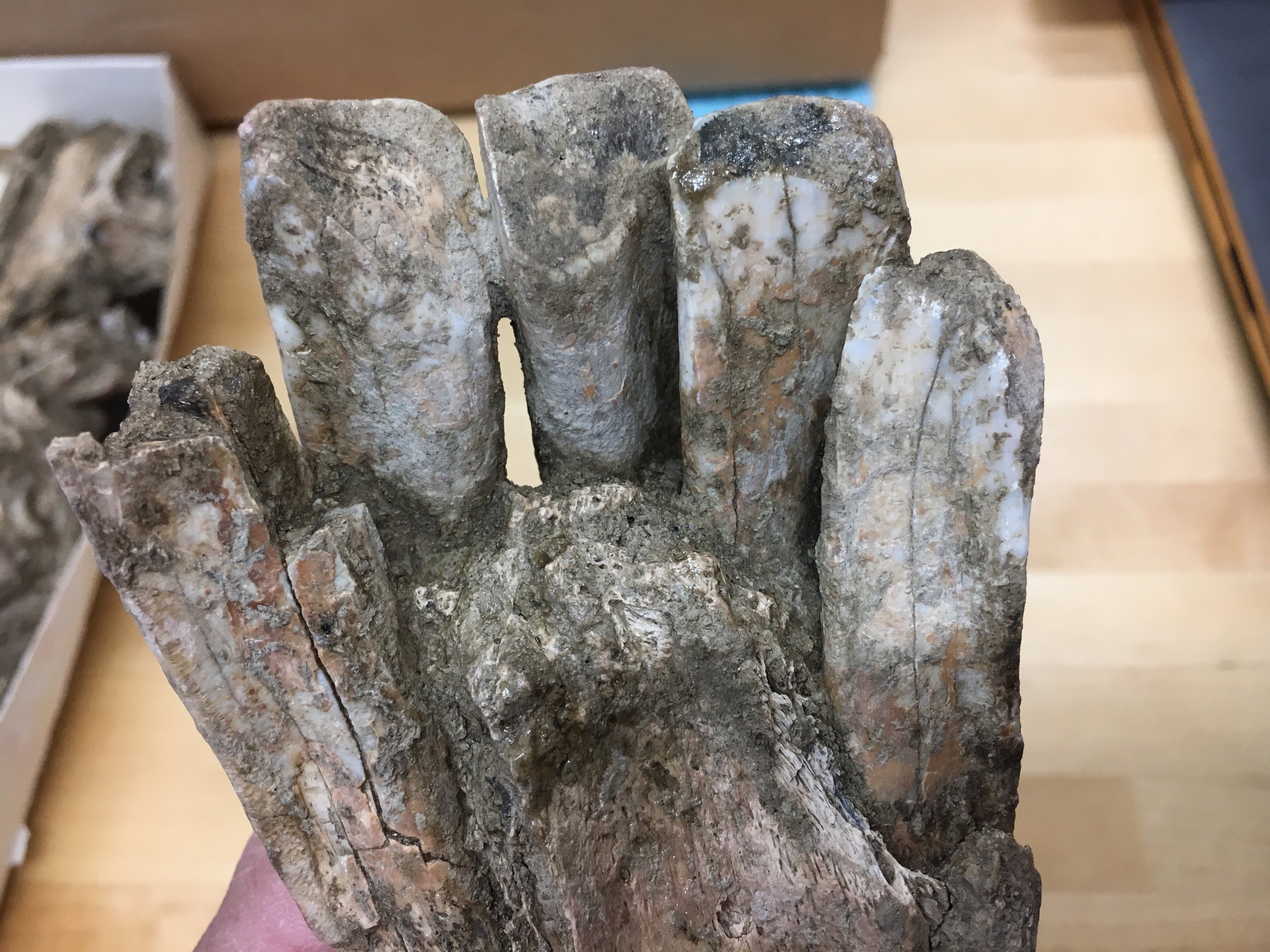 An interesting feature is that each of the three right incisors has a different shape and cross section. Using this specimen as a reference, and assuming there's not a huge amount of individual variation, we should be able to identify the exact position of isolated Camelops incisors from other specimens.We're working on making a 3D photogrammetric model of this specimen, which will be posted on our Sketchfab account in a few weeks.