

 I'm endlessly fascinated by the strange skeleton of the western camel, Camelops hesternus. Camelops was a massive animal, and many of its bones were so large they can easily be mistaken for those from a bison. Yet other parts of the skeleton are almost delicate.The long, slender bone shown above is a Camelops neck vertebra. Camelops had a long neck, but like almost all other mammals the neck had only seven vertebrae, so each vertebra had to be pretty elongate. (Most non-mammals with long necks have lots of shorter vertebrae, although there were exceptions.) The vertebra is shown in dorsal view (from above), and the front is to the right. The yoke-shaped structures at each end at the prezygopophyses (at the front) and the postzygopophyses (at the back), which articulated respectively with the zygopophyses of the preceding and following vertebrae.
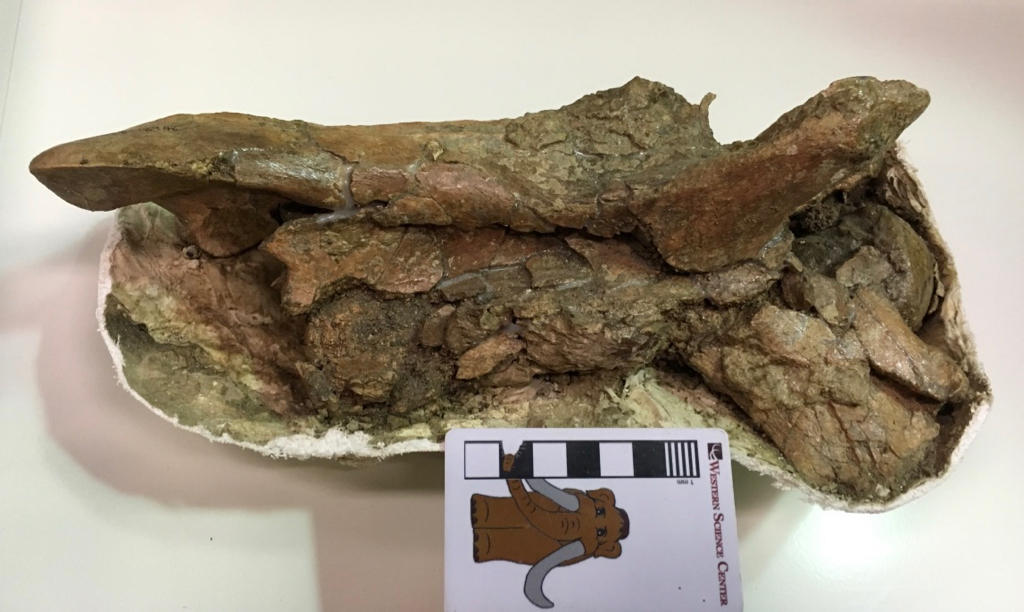 Looking at the right side, the ball joint at the front of the vertebral centrum is visible on the right. This corresponded to a socket on the back of the preceding vertebra, but didn't actually fit tightly into the socket as a large cartilage pad separated them in life. The large bony projection on the lower right is the right anteroventral costellar process. These processes provided attachment areas from some of the neck muscles, and are uniquely large in the camels.This particular vertebra was collected in 1999 near the East Dam of Diamond Valley Lake, and is probably around 50,000 years old.
Fossil Friday - Camelops jaw
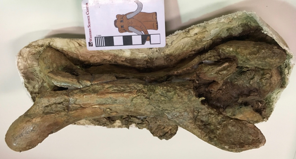
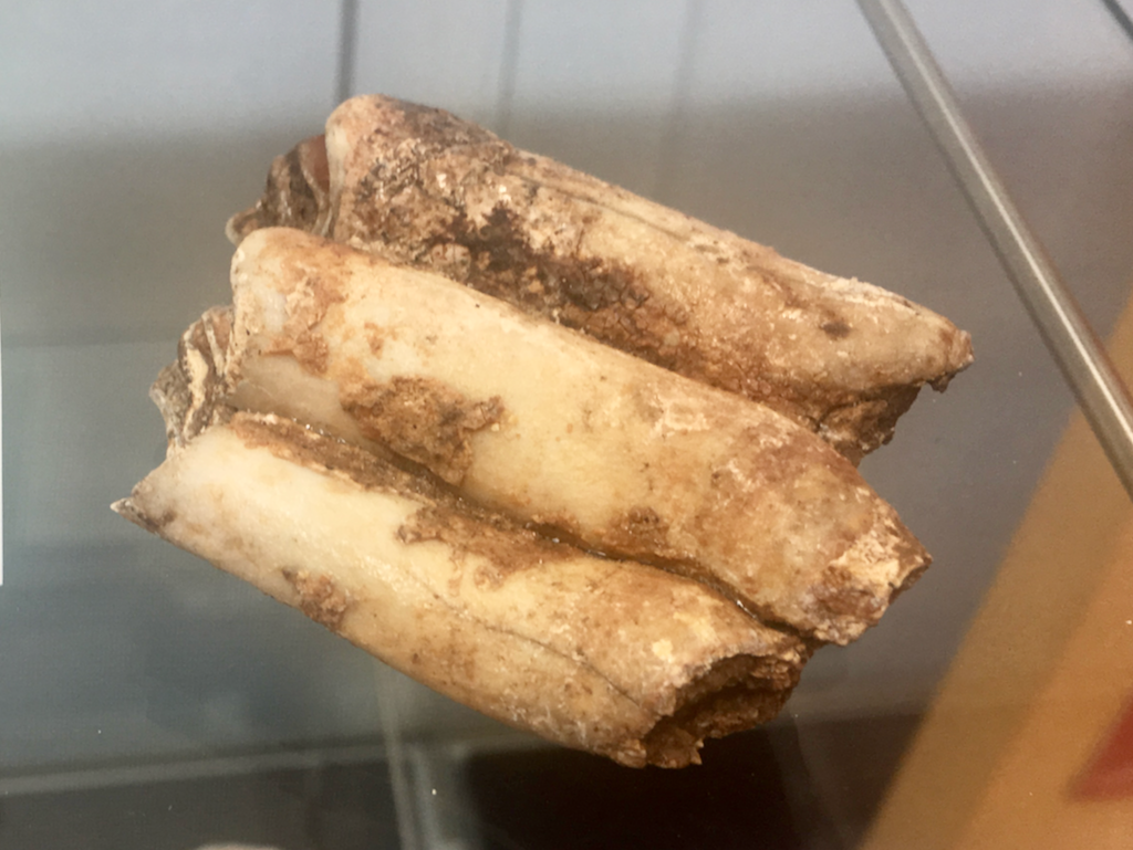
 This may look a bit like a zombie hand reaching from the grave in some B-horror movie (at least it does to me). In fact, it's a fossil that's quite a bit more interesting and less dangerous than a zombie hand.This is part of the lower jaw of the western camel Camelops hesternus, a fairly common species across western North America in the Late Pleistocene. This is the chin, technically called the mandibular symphysis, seen from underneath (ventral view). The forked structure on the left is behind the symphysis, where the left and right dentary bones separate to form each side of the lower jaw. The "fingers" on the right are the incisor teeth.Here's the dorsal view:
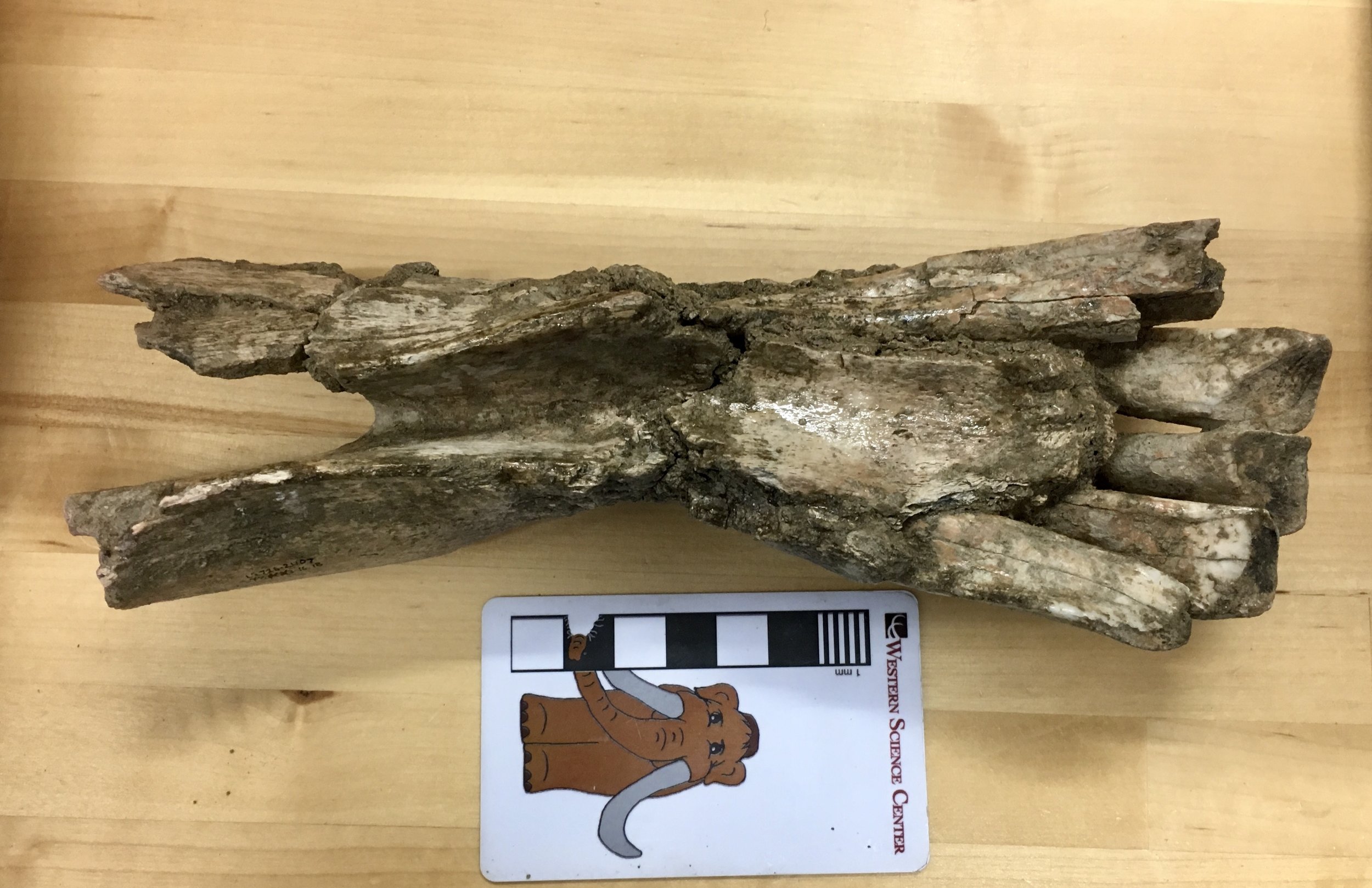 The incisors are more clearly visible in this view. Camels have three lower incisors on each side of the lower jaw; this specimen preserves all three on the right side, but only the first two on the left.Below is a lateral view of the right side:
 Because the bone is damaged on this side the entire length of the 3rd incisor is visible (minus whatever part of the tooth wore away before the camel died). Camels eat a tough, abrasive diet that is hard on teeth; camels compensate for this in part by having really long incisors that take a long time to wear down (the technical term - because OF COURSE there's a technical term! - is hypsodonty).We have more of this jaw, and it appears that there are no lower canine teeth. Since this is an adult animal, I suspect that it represents a female, since male camels (including Camelops) tend to have fairly prominent upper and lower canines. However, I need to do a little more research on Camelops to confirm that it displays this type of sexual dimorphism.Here's a close-up of the occlusal surfaces of the teeth:
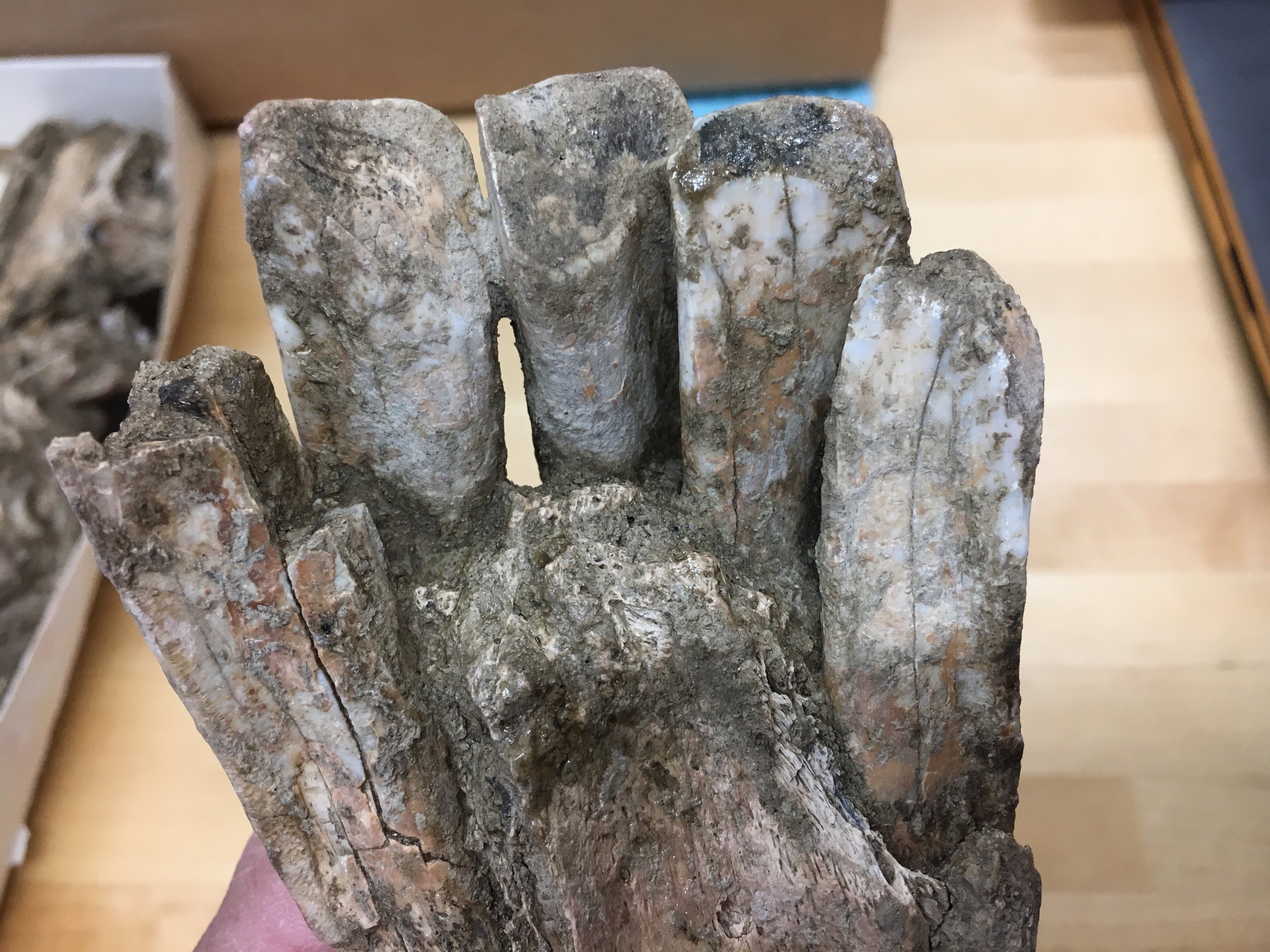 An interesting feature is that each of the three right incisors has a different shape and cross section. Using this specimen as a reference, and assuming there's not a huge amount of individual variation, we should be able to identify the exact position of isolated Camelops incisors from other specimens.We're working on making a 3D photogrammetric model of this specimen, which will be posted on our Sketchfab account in a few weeks.
Fossil Friday - Joshua Tree mammal bone
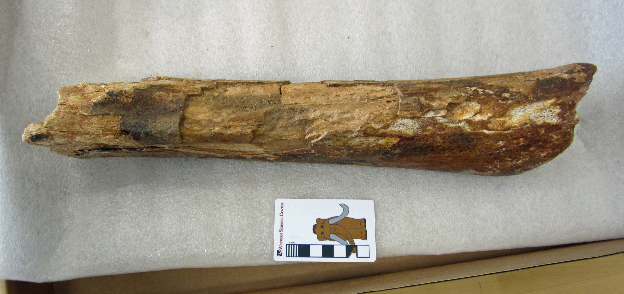 The Diamond Valley collection housed at WSC is an extremely rich record of Ice Age life in southern California, but it is far from the only Pleistocene site represented in the museum's collections.Right now, fossils of mammoths, camels, bison, horses, and other Ice Age megafauna from Joshua Tree National Park are on display in our "Fossils of Joshua Tree" exhibit. We are actively exploring new Pleistocene sites throughout Riverside County with colleagues from several institutions.In April, in conjunction with staff from the U.S. Bureau of Land Management, we collected this large incomplete mammalian limb bone from Pleistocene deposits near Blythe. It has recently been prepared in our lab, so now ahead is the task of comparing it to other large Ice Age mammal fossils and determining what kind of animal it is. It might belong to a large Ice Age camel such as Camelops hesternus, the species also found at Diamond Valley, La Brea Tar Pits, and many other sites in North America. We have much more discover in southern California's Pleistocene.Post by Curator Dr. Andrew McDonald.
Fossil Friday - Camelops tooth
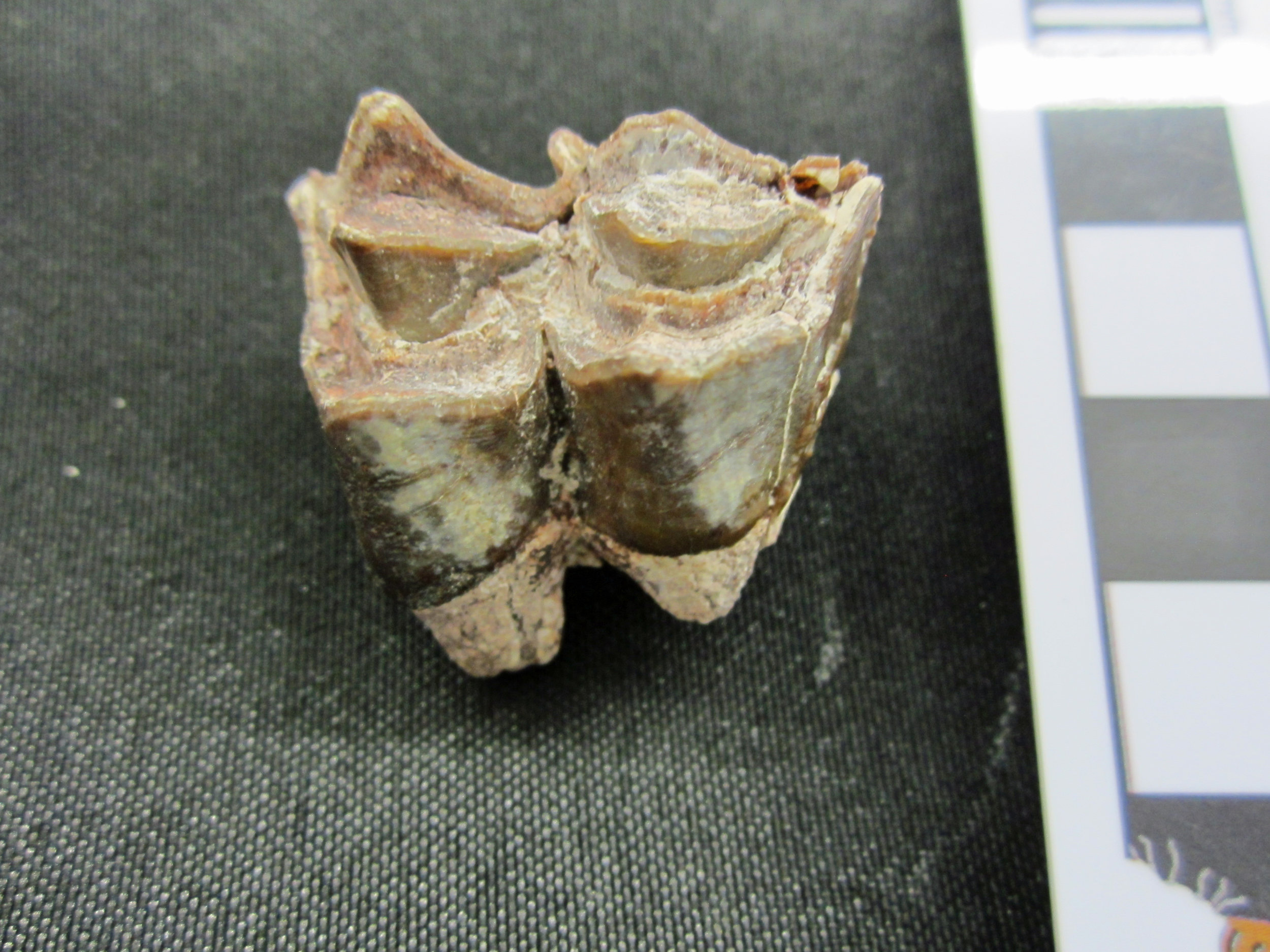
 Today's Fossil Friday specimen comes from the Pleistocene camel Camelops hesternus, a taxon we've featured several times on this blog. But this specimen is special because of where it was found - in Joshua Tree National Park.Joshua Tree NP is best known for its eponymous trees and spectacular granite exposures, but the park also preserves delicate desert habitats, native cultural artifacts, and fossils. A few months ago Western Science Center became a repository for Joshua Tree, and dozens of vertebrate fossils collected under the direction of Kathleen Springer were transferred here. One of the specimens is the tooth shown here, a lower right third molar from Camelops.To the extent that dating these fossils has been possible, they are roughly equivalent in age to the Diamond Valley Lake and Rancho La Brea fossils. Yet there appear to be real faunal differences distinguishing Joshua Tree from other localities, and there is a lot of exciting research ahead for these fossils.Another thing in their future is an exhibit. WSC is opening a new temporary exhibit, "Fossils of Joshua Tree National Park", which will include representative fossils from this collection. There is an opening reception for museum members tonight, and the exhibit will open to the general public tomorrow morning. Thanks to Vincent Santucci from the National Park Service, Melanie Spoo and the staff of Joshua Tree National Park, and Kathleen Springer of the USGS for making WSC the fossil repository for Joshua Tree and making this exhibit possible.
Fossil Friday - camel tooth
 Every day on my way to work, I drive past a farm that among its denizens counts several camels. Apart from the fun of seeing these large, strange mammals, they also serve as a reminder that wild camels once roamed across North America, until their extinction around 12,000 years ago. Although today, wild camels and their relatives are found in Asia, Africa, and South America, in a bit of evolutionary irony, camels actually originated in North America and spread around the world from there. The first camels appeared around 35 million years ago during the Eocene Epoch, in the form of small fast-running herbivores like Poebrotherium. By the Miocene Epoch, camels had diversified and grown larger. This image shows one of the upper left cheek teeth of a camel that lived here in California between 18 and 14 million years ago. This tooth was collected by the Western Science Center earlier this year at a fossil site in San Bernardino National Forest. It might belong to an extinct camel called Aepycamelus, which was widespread in North America during the Miocene. However, we are still studying this tooth to determine exactly what species it represents.
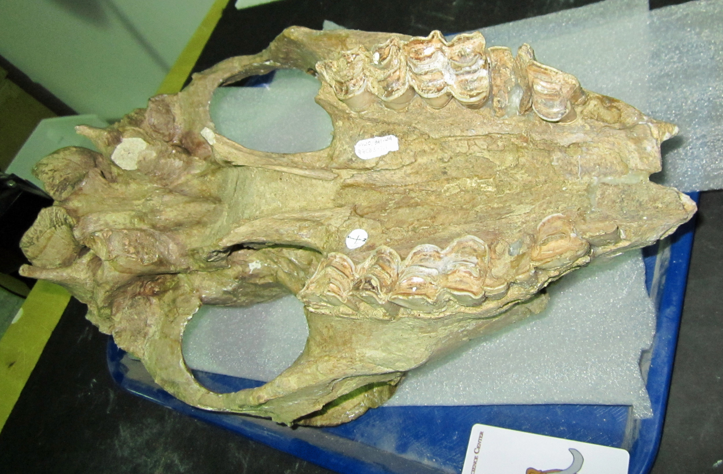 Camels kept going strong in North America up until their extinction at the end of the last Ice Age. One of the last camels in North America was the large species Camelops hesternus, which ranged across western Northern America, including California. This image shows the underside of a Camelops skull discovered at Diamond Valley Lake and which is part of the Western Science Center's collection. The teeth are highly worn, but still bear a close resemblance to the smaller Miocene tooth. Post by WSC Curator Dr. Andrew T. McDonald
Fossil Friday - camel elbow
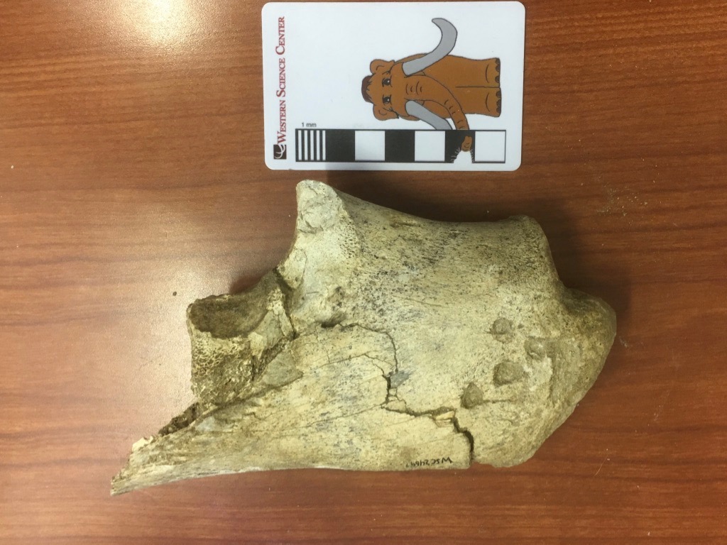 We're continuing our focus on Pleistocene fossils from Murrieta, California this week with a single bone fragment that has a lot going on.This bone is labeled in our collections as an ulna from Equus, a horse. It is indeed part of a left ulna, one of the bones from the forearm. More specifically, it's the olecranon process, the proximal end of the ulna that forms the point of the elbow. The triceps muscle attaches to the olecranon process, allowing you to straighten your arm (or front leg, in this case). However, after spending several hours comparing this to various publications and our Diamond Valley Lake collections, I'm not convinced it's a horse.For an olecranon process of this size, there are really only three likely animals from the Pleistocene of Southern California it could belong to: horse, camel, and bison. Everything else is either much larger (mammoth, mastodon) or much smaller (mule deer). (OK, to be fair, short-faced bears and ground sloths are in this size range, but their ulnae look nothing like this.) Horses, camels, and bison are all known from this site, but while it's a little on the small side, this bone is the best match with the western camel, Camelops hesternus.There is another interesting feature on this bone. Notice the four circular depressions near the tip (on the right). Here's a closeup:
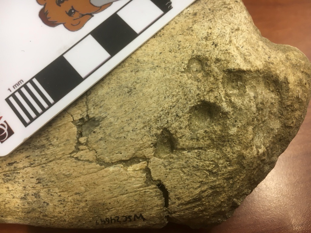 There are a few comparable depressions on the other side:
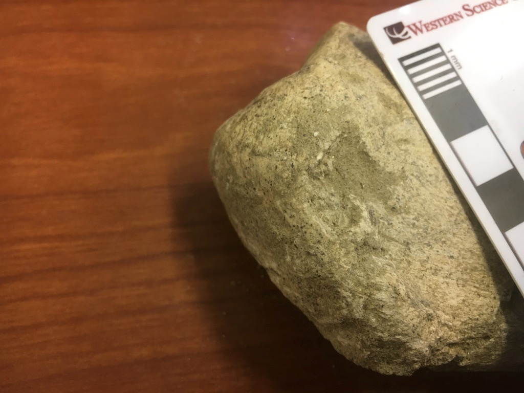 These appear to be bite marks, from some carnivoran chewing on the end of the bone. There are several other scrapes that appear to be gnaw marks. So far we have not identified any carnivoran bones from this site, but they certainly made their presence felt.We have scanned and 3D-printed this bone (print shown below with the original), and the scans can be viewed on Sketchfab at https://skfb.ly/6xNMM.
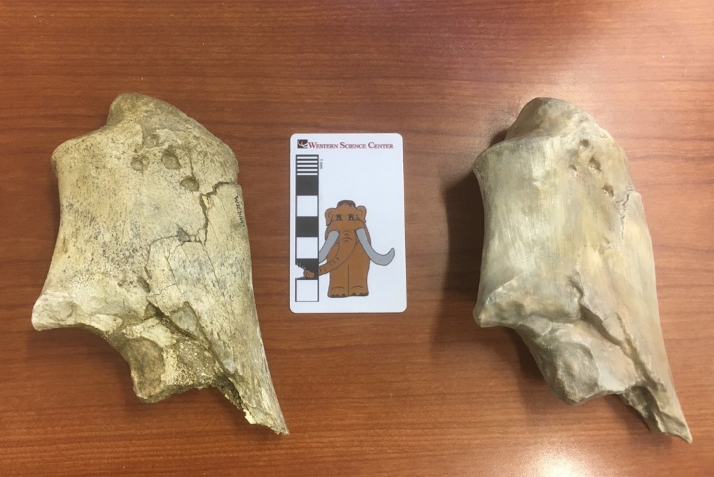
Fossil Friday - camel vertebra
 This is a cervical (neck) vertebra of a giant extinct camel called Camelops, which roamed southern California during the Pleistocene Epoch, perhaps less than 50,000 years ago. This particular specimen was discovered in 2002 near Murrieta, and is part of a fauna that also includes horses, mammoths, and giant ground sloths. The view shown here is the right lateral view of the vertebra, showing the ball-shaped structure and large prongs (prezygapophyses) that would have articulated with the next vertebra closer to the head.
Fossil Friday - camel lumbar vertebra
 While we only have one well-preserved skull of the extinct camel Camelops hesternus from Diamond Valley Lake, we have a large number of post-cranial remains.The bone shown above is a lumbar vertebra, seen in anterior view. This seems to be the 7th lumbar, the last one in the series before the sacrum (which was also recovered from this individual, along with several other bones). The prominent curved structures above and on each side of the neural canal are the prezygopophyses. These articulated with the postzygophyses of the 6th lumbar. The strong curvature would largely lock the two vertebrae together, resulting in a relatively inflexible lumbar region.Here is the posterior view, with the postzygophyses visible:
 And the left lateral view:
 The neural spine is broken on this specimen, as are the transverse processes (the right one is missing entirely). The broken surfaces are packed with sediment and abraded, indicating that they were broken off before burial. That is interesting considering that there are multiple associated bones with this specimen; that makes it less likely that the bone was damaged by, say, washing down a river. Could this damage have been caused by scavenging? I tried looking at the bone with low-angle light, which sometimes helps reveal bite marks on the surface:
 Sure enough, these are apparent bite marks on the bottom edge of the centrum. Below is the preserved part of the left transverse process, with apparent bits marks along its entire length:
 There are other possible bite marks scattered across this vertebra. Moreover, close examination also revealed possible insect feeding traces in various places, including on one of the prezygapophyses:
 These possible traces seem to be extremely common on bones from Diamond Valley Lake, possibly occurring on half or more of the large specimens. Clearly, besides studying the bones themselves, there's a lot of potential in the DVL trace fossils as well.
Fossil Friday-camel humerus
 In spite of the facts that camels are among the more common large animals from Diamond Valley Lake and are intrinsically cool, I've somehow managed to get almost halfway through 2016 without featuring them on Fossil Friday. I'll rectify that today.Above is a left humerus (upper arm) from Camelops hesternus. It's shown anterior (or cranial) view, with the proximal (upper) end on the left. The profusion at the lower left corner is part of the humeral head, the "ball" part of the "ball-and-socket" joint at the shoulder, while the articulation for the radius and ulna (the elbow joint) is on the right.This bone is still in its field jacket, and like many of the large bones from Diamond Valley Lake has only been partially prepared. Slowly but surely we're going to be finishing the preparation work on these bones, but it's a process that will take many years.
Fossil Friday - wildfires
 A large wildfire, called the "Lake Fire", is currently burning in the San Bernardino National Forest. Even though the fire is about 50 km north of Hemet, smoke from the fire is clearly visible from the Western Science Center.Wildfires such as this are widespread in Southern California during the summer, driven by dry vegetation and frequent afternoon and evening winds. The regularity of these fires is evident in the fleet of Cal Fire aerial tankers based at the Hemet Airport, which are currently making regular flights to combat the Lake Fire:
 Anthropogenic climate change and the ongoing drought have resulted in ideal conditions for wildfires in this area, but wildfires are not a new occurrence in California. In fact, a quick records search of the Diamond Valley Lake fossil collection housed at the museum turned up at least 80 Pleistocene specimens that show evidence of burning. The DVL fossils all predate the arrival of humans in California, so these aren't animals that were cooked for food. They represent animals that were exposed to wildfire at or fairly close to the time of death.In some cases the evidence for burning is subtle, but in others there is no room for doubt:
 Above are two views of the left tibia (shin bone) of the camel Camelops. Most of the bone is missing, with only the distal end preserved (there is also a box full of associated fragments). The bone is completely burned, inside and out, and has almost a charcoal-like texture on the surface. The burning extends to the broken surface, so presumably the bone was broken when it burned. It's possible that the bone had been exposed on the ground for awhile and had already started cracking up when the fire came through, but it's also possible that a relatively fresh bone cracked and broke due to the heat from the fire. Regardless, specimens like this show that, much like today, wildfires were a regular occurrence in Southern California during the Pleistocene.
Fossil Friday - bite marks on a camel skull
 One of the specimens we have on display at the Western Science Center is a cranium and partial vertebral column including the neck of the camel Camelops hesternus. A closer examination of the skull reveals some surprising features. The parietals (the bones that make up the back half of the top of the braincase) have a series of holes and apparent scrapes:
 At first I thought the two back holes might be an anatomical feature called the parietal foramen. However, parietal foramina are uncommon, usually forming as a developmental abnormality. I've been unable to find images or reports of parietal foramina in Camelops or any other camel. The holes in this specimen are not symmetrical in their position (each one is located in a different position on its respective parietal), and there are cracks in the parietals leading to the holes. Finally, if these were parietal foramina, it doesn't help to explain the presence of the other hole located at the front of the parietal. Taken together, this suggests that the holes are not an anatomical feature but instead are bite marks from another animal.I originally envisioned a carnivore grabbing the camel by the top of the head, with its head held almost parallel to the camel's head, and then dragging the camel's head and neck away from the rest of the carcass. This would mean that the four parietal marks (three punctures and one scrape) were made by the four canines of the carnivore in a single bite. Darla and I pulled out cast skulls of several different carnivores to try this out, but we couldn't get it to work. If we assume that the four marks were made in a single bite, then the spacing is about right for a small dire wolf, a black bear, or a jaguar. However, when we tried to simulate the bite the predator's incisors would always hit the sagittal crest (the ridge of bone along the top of the skull) long before the canines reached the parietals. If we went with larger animals with longer canines like a short-faced bear or an American lion, the canines would reach the parietals but the spacing was far to close for such a large animal.I now think that it's more likely that the punctures were caused by multiple bites, by a carnivore coming at the skull from an oblique angle (possibly multiple angles), something like this:
 This actually may be more consistent with the behavior of modern carnivores; if you want gory confirmation, do an image search for "hyena carrying head" for examples of how scavenging carnivores transport prey. There is also some evidence that there may be additional bite marks all over this skull, consistent with multiple bites. There are two unusual depressions in the top of the frontal bone, several holes along the edge of the right lambdoidal crest at the back of the skull, and damage to part of the right squamosal. To be fair, none of these additional marks are sure things; if not for the four bite marks on the parietal I would have never considered these as likely candidates for bite marks.Another observation I would not have thought twice about except for this context is the damage at the front of the skull. The nasal bones are damaged, and the premaxillary bones are missing entirely. The premaxillae can loosen and fall off as a skull dries out, and that's normally how I would explain this. But if this skull was being chewed up by carnivores it raises another possibility. It seems that the nose is one of the choice bits of meat on the skull for a carnivore. There's a lot of meat and blood in the nose, and it's easier to get to than the brain. Moreover, while cats such as lions usually kill their prey with a bite to the neck, closing the windpipe, an alternate method used on occasion is to bite down on the nose. Is it possible that the missing premaxillae in this specimen is another predation feature?
Fossil Friday - camel molar
 For this week's Fossil Friday we'll return to camels, specifically the large extinct camel Camelops hesternus that's pretty common in Pleistocene deposits in California.This specimen is an upper molar collected near the west dam of Diamond Valley Lake. I'm pretty sure this is the upper right first molar, although I can't yet rule out the possibility that it's the second molar. This tooth was found associated with several other molars and premolars, as well as small skull fragments that all appear to be from one individual.The image above is in occlusal view, showing the chewing surface. The tooth is fairly heavily worn, showing the pattern of folded enamel ridges; the shiny grayish-white ridges are enamel, with softer dentine in between. By having these alternating areas of hard enamel and softer dentine, the enamel ridges always stick out slightly beyond the occlusal surface, so the tooth maintains a sharp chewing surface even as it's being worn down. The elevated enamel ridges are especially noticeable in lingual view (since this is an upper tooth, the occlusal surface is at the bottom):
 The particular pattern of enamel ridges is one of the primary means of distinguishing between different groups of mammals, even down to individual species. For some species it's possible to make an identification on the basis of a single well-preserved tooth.Camels have a general tooth pattern that's called selenodont, in reference to the crescent-shaped enamel ridges in occlusal view. Camels share the selenodont pattern with many of the camel's artiodactyl relatives, including cervids (deer) and bovids (bison, cows, antelopes, etc.).Just for the sake of completeness, here's the labial view of the same tooth:

Visiting the Raymond M. Alf Museum
 I spent today continuing to familiarize myself with Southern California by visiting the Raymond M. Alf Museum of Paleontology, located in Claremont on the campus of The Webb Schools. Once I arrived, Museum Director Don Lofgren, Curator Andy Farke, and Outreach Director Kathy Sanders kindly spent the better part of a day showing me around their museum and discussing museum operations and California paleontology.The Raymond Alf Museum is operated by The Webb Schools, a private high school, and paleontology and museum operations are heavily integrated into the school's curriculum. Many students participate as authors on research projects, including the description of "Joe", a baby Parasaurolophus published last year in PeerJ, and now on exhibit in the museum:
 Of course, the Western Science Center also shares a campus with a school, the Western Center Academy. One of my goals in visiting the Alf Museum is to see how they've combined their research and educational efforts.Besides "Joe" there are tons of interesting fossils and casts on display in the museum. There are numerous titanothere skulls from Eocene deposits in South Dakota and Nebraska:
 The Alf has one of the most impressive displays of fossil vertebrate trackways I've ever seen. There are plenty of dinosaur tracks, but how many museums have multiple examples of fossil camel tracks?


 Or how about cats?
 There are also numerous impressive slabs of Permian Coconino Sandstone that are covered with multiple reptile trackways:
 I'd like to thank Don, Andy, Kathy, and the rest of the staff at the Raymond Alf Museum for taking the time to meet and show me around their excellent museum.
Fossil Friday – more camel bones
 Last week for Fossil Friday I showed an example of a metapodial of an extinct camel, Camelops hesternus, which was collected about a mile from the museum's current location. It turns out that the metacarpals weren't found in isolation. Several other bones were found nearby, including the two large fragments shown above.These relatively large fragments, like the metapodial from last week, are both camel bones. But we've moved up the arm; these are fragments of the humeri (upper arm bones). In each case, only the end near the elbow (the distal end) is preserved. I've photographed them from the front (cranial or anterior view), in the same orientation as you would see them in a complete skeleton. That means that the fragment on the left is actually the right humerus, and the larger one on the right is the left humerus. Almost half the left humerus is preserved. Compare it to this much more complete specimen from the WSC collection (seen in lateral view, with the distal end on the left):
 It's possible that the two humeral fragments and the metapodial from last week belong to the same individual camel, especially since there were additional bones from the front legs associated with these. However, at this point I can't say with certainty that they come from one animal. I haven't yet seen the original field notes or photos, so I don't know if they were found actually articulated with each other or if they were just nearby, but I do know that there were bones from other species found at the same site. It would be worthwhile to measure each camel bone and compare their proportions to known associated bones of Camelops to see if their relative sizes are consistent with one animal, and to check them for indications of whether or not they're at the same growth stage. These checks won't prove that they come from one animal, but depending on the results they could prove that they don't come from one animal.Finally, another point is worth noticing. The second specimen is only partially prepared, with one side still in the original field jacket. A large number of specimens in the WSC collection have been prepared only enough to make an identification, and still need to be fully cleaned and restored. We have a lot of work to do!
Fossil Friday – camel metacarpal
 For Fossil Friday, we have the hand bone (front foot bone) of the western camel, Camelops hesternus, seen here in anterior view (the bottom is to the left). It was collected about a mile from where the museum is now located, and was associated with several other camel bones.Like many other members of the Artiodactyla, the "hand bone" (or metapodial) in camels is actually two bones fused together, the 3rd and 4th metacarpals. Even though the two bones are fused together for most of their length, there is still a visible groove indicating where they're fused, and at the distal end the bones are still separated. At the distal end each metacarpal articulates with a separate finger; the articulation is more clearly visible in the posterior view of the same bone, with the articulation on the left:
 These correspond to the middle and ring fingers in humans. The foot has a similar arrangement. Since most artiodactyls have this two-finger, two-toe arrangement they are sometimes called the "even-toed ungulates".It's easy to forget how big camels are. Camelops hesternus was roughly the size of modern camels, and they are massive animals. This metapodial is close to 35 cm (over a foot) long. Here's what a whole skeleton looks like, from the exhibit at the George C. Page Museum:
 Even though Camelops hesternus was comparable in size to a modern camel, it was actually more closely related to the llamas and vicuñas from South America, and was the largest member of that group. Camelops went extinct shortly after the end of the last Ice Age.