by Outreach & Communications Coordinator Brittney Elizabeth Stoneburg
Last week the Western Science Center released a new paper about Micoene horses from Southern California in UC Berkeley Museum of Paleontology’s open access journal PaleoBios. This research has given us a clearer picture of what the area around San Bernardino National Forest looked like millions of years ago, and all it took was a few small horse teeth.
Excavations have been going on in the Cajon Valley Formation for years, but in 2018 Western Science Center researchers were granted a permit by the U.S. Forest Service. It dates back to the Miocene Epoch, which spanned 23.03 to 5.333 million years ago, and the Cajon Valley Formation fossils in the WSC collections are approximately 16.5 to 14 million years old. Horses living during this time period were much smaller than modern horses (a newspaper article on paleontologist John Merriam’s excavations nicknamed them “doll ponies” in 1913) and had three toes on each foot, unlike modern horses, which only have one toe on each foot.

The field site in 2018, with specimen positions marked. Lead author Brittney Elizabeth Stoneburg for scale.
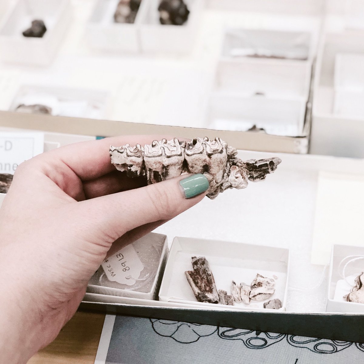
We eventually identified three species of horses from the WSC sample - Archaeohippus mourningi, Scaphohippus sumani, and Parahippus brevidens (or as I tend to refer to their respective sizes in my head, tall, grande, and venti!). Each of them helped us gain a clearer understanding of prehistoric California, and what the environment there was like millions of years ago.

Teeth from the WSC Cajon Valley Formation sample. A. Parahippus brevidens. B. Archaeohippus mourningi. C. Scaphohippus sumani.
While it wasn’t particularly surprising to have found several Ar. mourningi specimens, as these tiny horses had been reported before in the formation, Scaphohippus and Parahippus ended up being a more complicated story.
Scaphohippus, for example, was originally separated into two species - S. sumani and S. intermontanus. As mentioned, you can identify many different species of horse based primarily on their teeth, and Scaphohippus is no different. S. intermontanus was said to have a less complex tooth surface than S. sumani, and was mostly known from the nearby Barstow Formation. But as we visited museum collections and tried to determine which species of Scaphohippus we had in our sample, we grew increasingly confused. These horse teeth looked so similar that it was incredibly difficult to tell the two species apart! Finally, instead of comparing individual teeth, we decided to look at whole tooth rows, and that sparked a question.
We had just assumed that the idea of there being two species of Scaphohippus was correct, but what if it wasn’t? What if the reason the teeth looked so similar is because they came from the same species?

How the realization felt to the authors.
Many of the fine details that help us identify horse teeth appear with a certain amount of wear, or in a particular tooth position. In Scaphohippus, the defining features are called “plications”, small enamel folds in the center of the tooth that increase in number as the horse would wear the tooth down with use. The number of plications in a particular tooth was part of what differentiated S. sumani from S. intermontanus, but when we looked at the tooth rows, we realized this difference wasn’t consistent enough to base an entire species on. And so our question was answered - our specimens belonged to S. sumani, because we no longer considered S. intermontanus a separate species! (S. sumani was named first, and so that name had what’s called “taxonomic priority”).
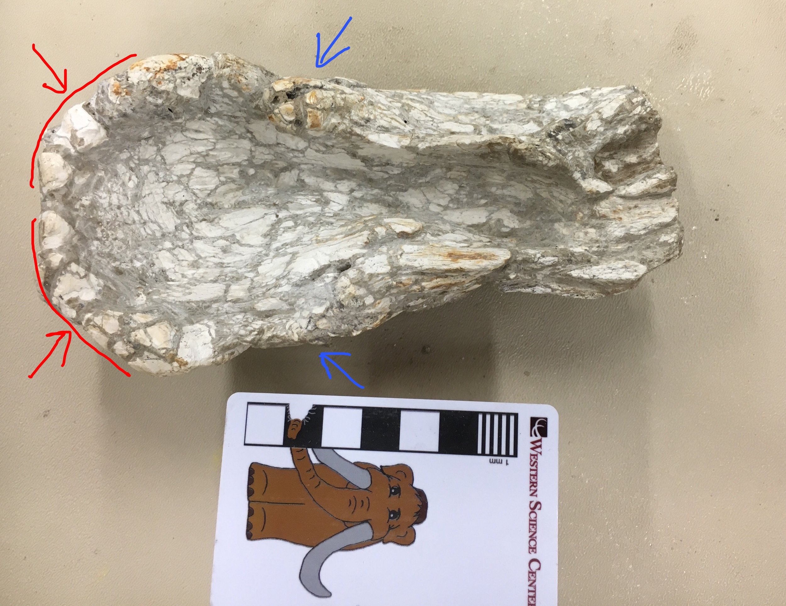
After that, it was time to turn our attention to a particularly strange little tooth that didn’t fit either Archaeohippus or Scaphohippus. Shaped like an ear lobe, this molar was markedly different from all the other teeth we had collected.
We searched high and low for an identification, but no previously published specimen from the Cajon Valley Formation matched. Then, at last, we stumbled on Parahippus brevidens, a Miocene horse species that had first been identified in the Mascall Formation in Oregon. Our tooth matched the holotype (the fossil a species’ description is based on) almost exactly.
Parahippus brevidens had been mentioned here and there in databases with Cajon Valley Formation specimens, but this paper is the first research confirming that these horses did indeed live in Southern California. This has extended the known range for these animals by about 400km and means they appeared up and down the west coast, at least from Oregon to Southern California! We learned so much new information about this horse species and its range, all from a single tooth.
So we figured out what three species of our horse we had in our sample - great! But what does that mean?
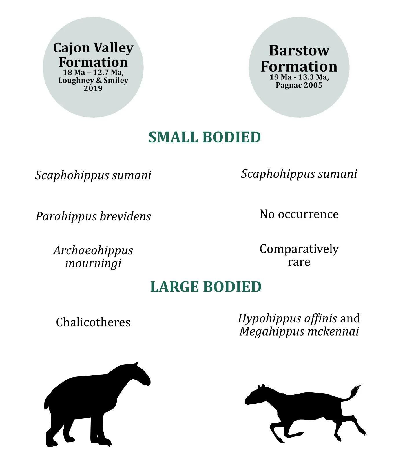
A faunal comparison between the Cajon Valley Formation and the Barstow Formation. Adapted from Figure 7 of Stoneburg et al (2021).
It means that this research is another piece in the puzzle of figuring out what California's ancient past looked like. Knowing now that we have three species of horses in the same place gives us a clearer picture of the Cajon Valley Formation's ecological profile. For example, we can now more accurately compare and contrast the formation’s fauna with the well-studied Barstow Formation, which is similar in age to the Cajon Valley Formation and is only around 65 miles away. Yet we see these consistent differences in the fauna found in each formation - for example, while Parahippus brevidens is now confirmed to have been found in the Cajon Valley Formation, it has yet to appear in the Barstow. Larger horses, like Hypohippus affinis and Megahippus mckennai, appear in the Barstow Formation, but haven’t appeared in the Cajon Valley Formation. What caused these faunal differences? Was there some sort of geological barrier impeding movement? Were the environments markedly different despite the proximity? We don't know quite yet, but these are the types of questions we can start asking.
There’s still so much we don’t know about the Cajon Valley Formation, and California during the Miocene as a whole, so every bit of research helps expand our understanding of a long gone environment. You can definitely expect more research about the “doll ponies of Southern California” to come!

 For my first research project, I've been working on fossils collected by the Western Science Center from our field site in San Bernardino National Forest. This means I'm up to my neck in horse teeth right now! I've been focusing on the small, three toed horses that roamed Southern California during the early to middle Miocene, approximately 18 million years ago.
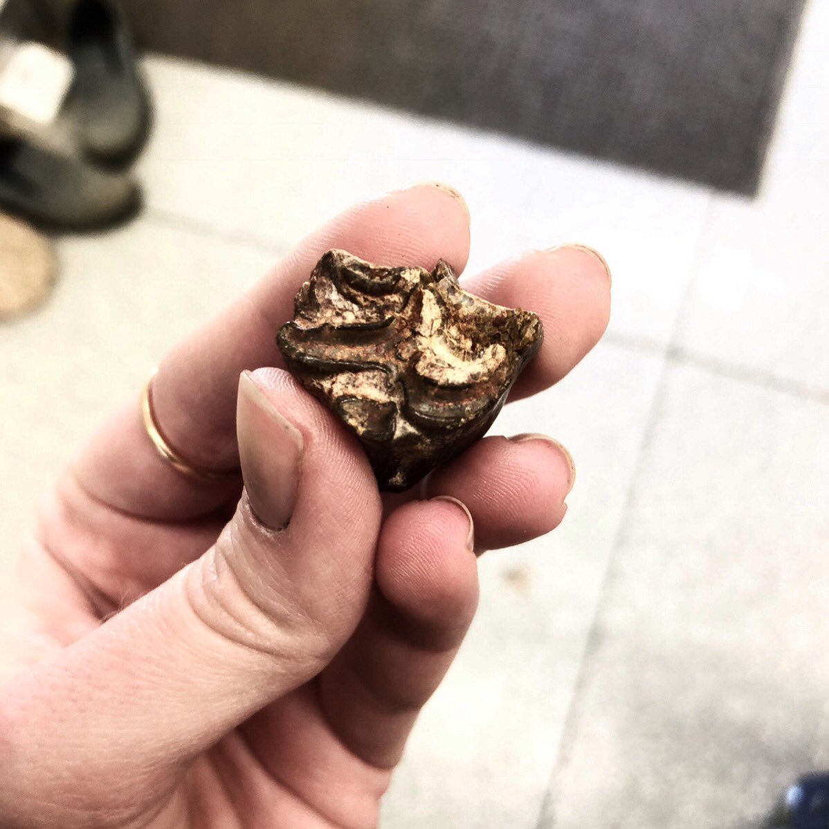 Multiple species of horse lived in this area, including Scaphohippus, whose teeth are picture above. While our collection from this site has only a few post-cranial bits, we can make pretty solid identifications just from the teeth. Horse teeth like these have numerous features that make it possible to identify them down to the species level. Knowing what horses lived in this area during the Miocene can tell us a lot not only about the horses themselves, but about the environment they lived in!I'll be presenting on my research at the
 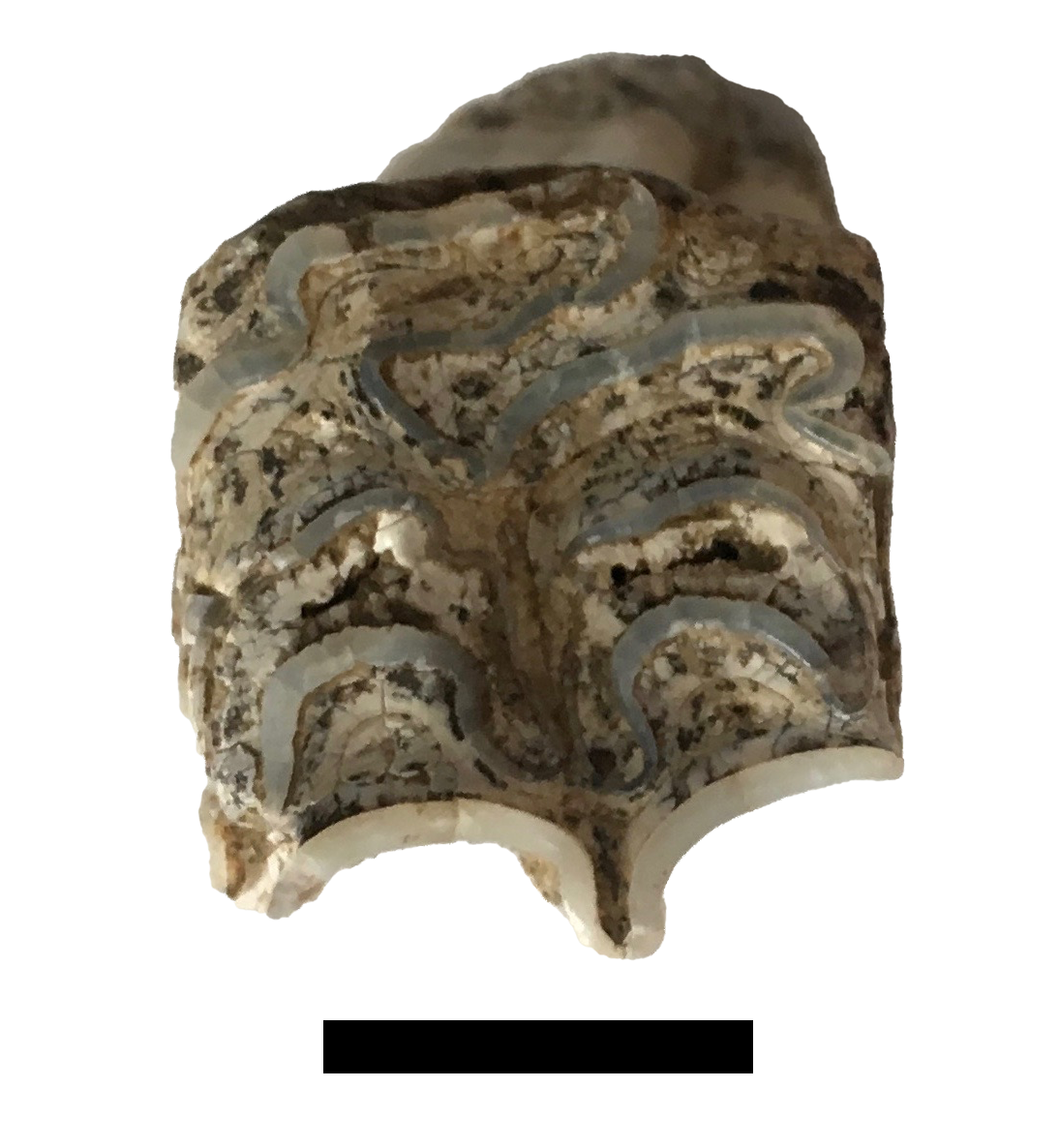 One thing has become quickly obvious as we've been examining the Harveston fossil collection: there are a lot of horses.This is one of several isolated horse teeth from Harveston. This specimen is an upper right 1st molar, and is of particular interest because it is quite a bit smaller than most of our Harveston horses, as can be seen below:
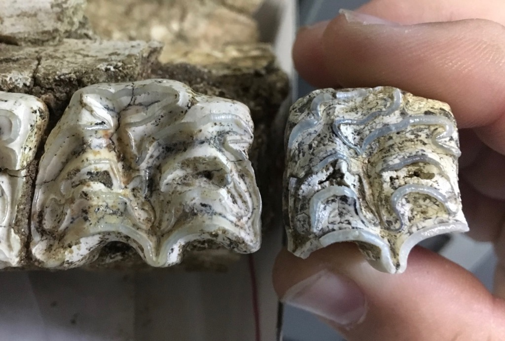 It turns out there were actually two species of horses at Harveston. The larger and much more common one is Equus occidentalis, while the smaller, rare species is generally called Equus conversidens (there are some nomenclatural issues with this name and with Equus species in general, but I'm not going to get into that here). E. conversidens makes up perhaps 5% of the Harveston horses. This seems to be a general trend for southern California sites, including Diamond Valley Lake, with both species generally present, and E. occidentalis being much more common.We've made a 3D scan of this E. conversidens tooth (WSC 24676) available for download on Sketchfab at
 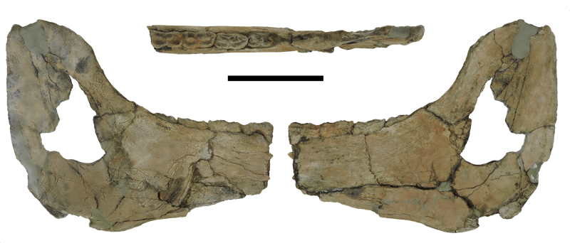 Fossil Friday this week continues our examination of Pleistocene specimens from Temecula Valley, this time with a partial lower jaw from the horse Equus occidentalis.The image above was prepared for our poster at next month's regional GSA meeting in Flagstaff. It shows the dentary in medial, dorsal, and lateral views. The scale bar is 10 cm. In the dorsal view, you can see the occlusal surfaces of the last four teeth in the jaw, which in this case are (from front to back) the 4th deciduous premolar, and molars 1, 2, and 3. The 3rd molar had only just started to erupt, suggesting that this horse was about 3.5 years old when it died. In an oblique anteromedial view (actually a screenshot from a photogrammetric model), you can see the unerupted 4th premolar underneath the 4th deciduous premolar, and, in the background, the deep crown of the 2nd molar where the side of the jaw is broken:
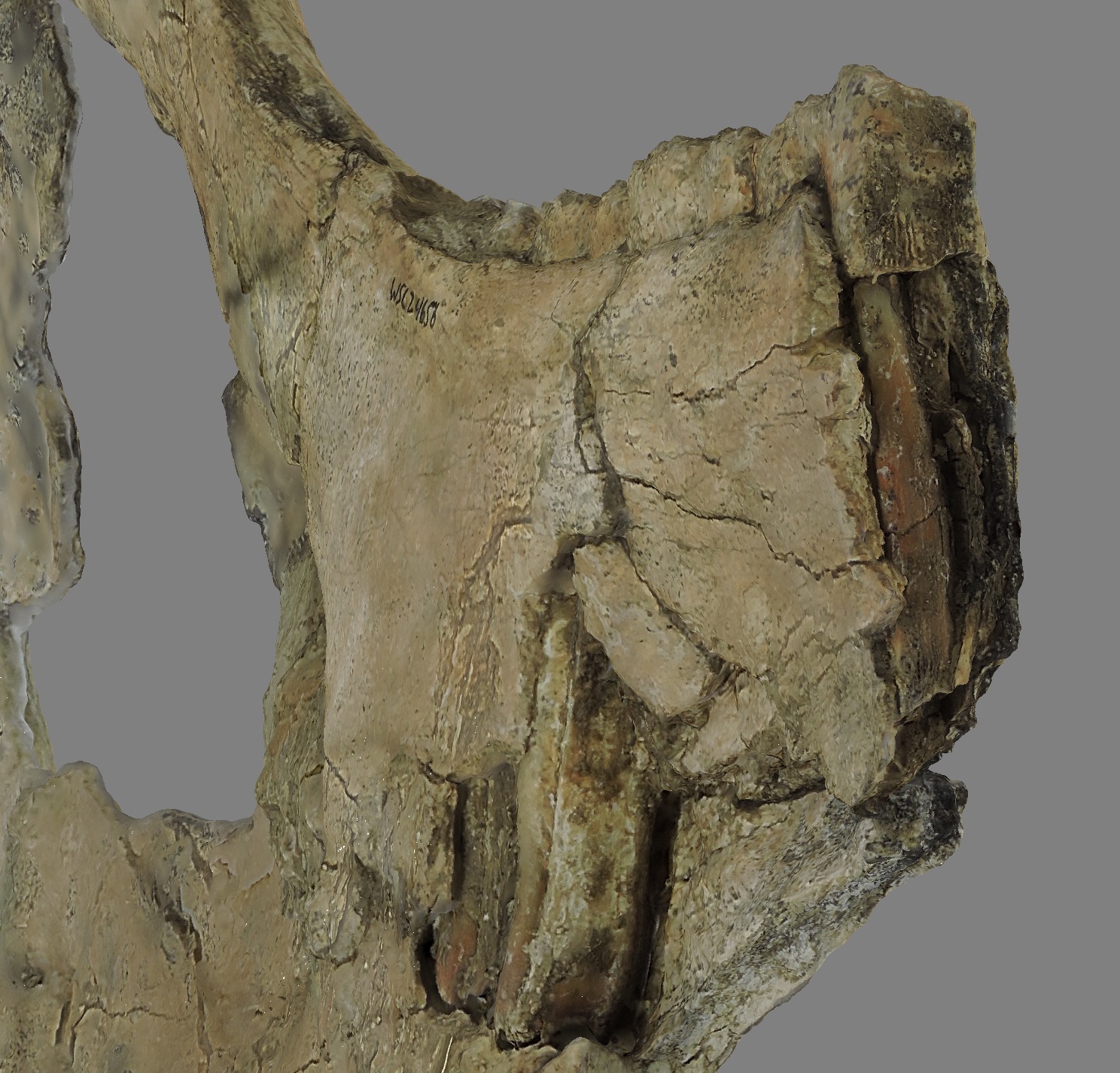 We've made a photogrammetric model of this specimen, and printed a 3d replica that we've already started using in programming:
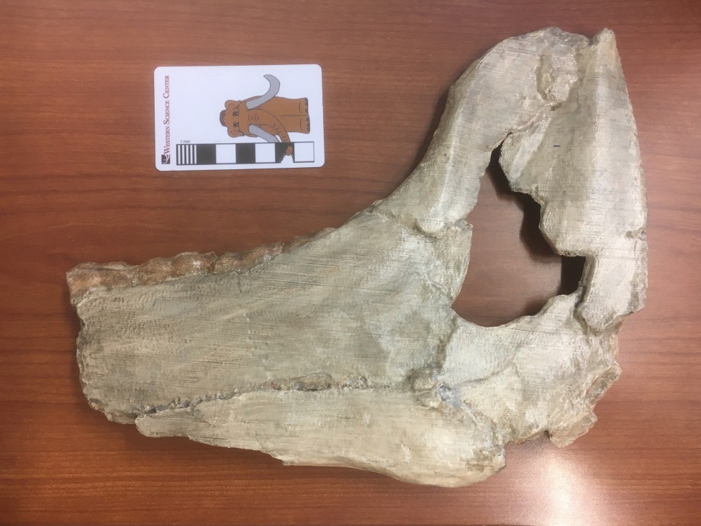 The 3D file of this specimen is available for download at WSC's Sketchfab site at
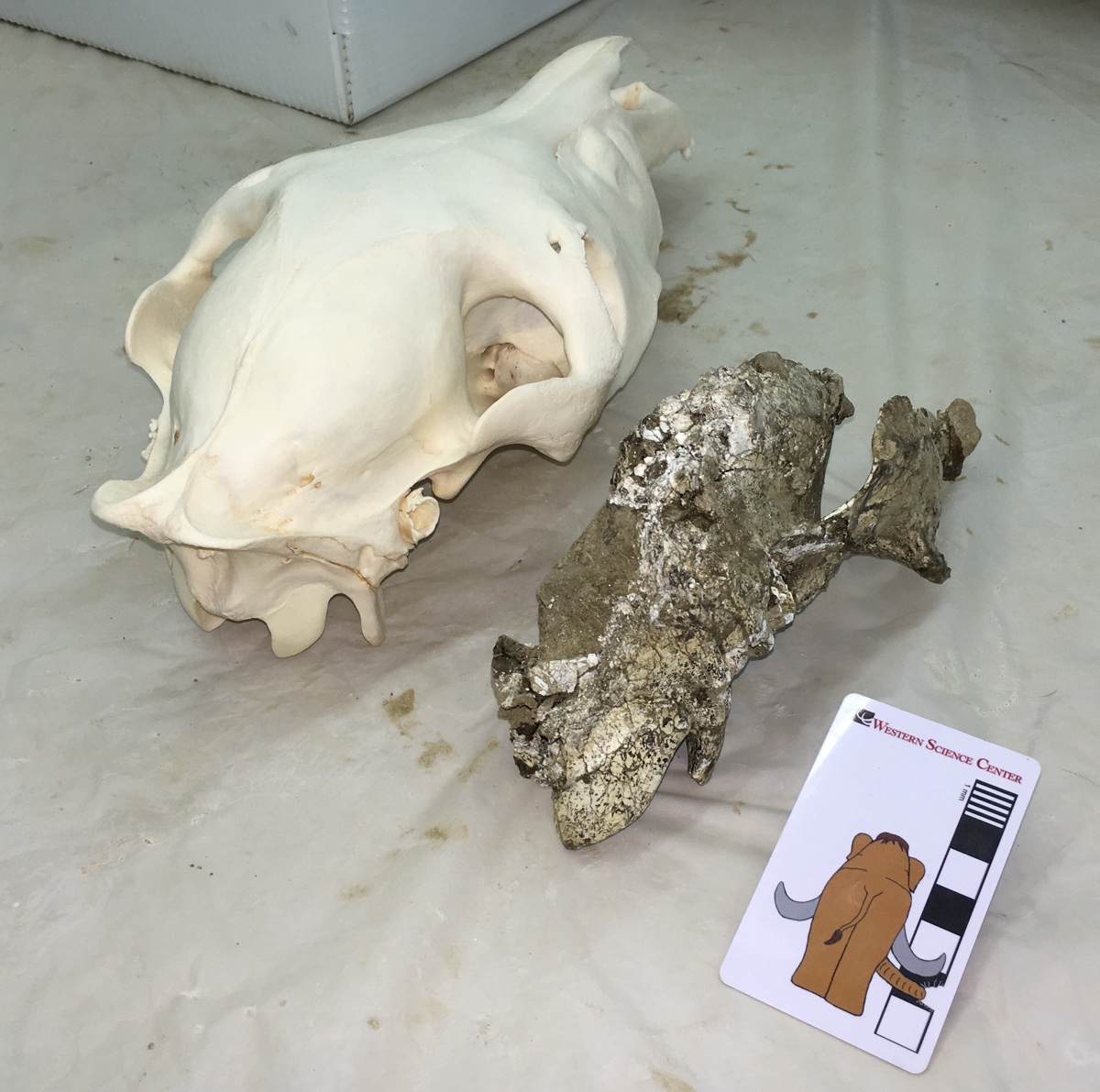
 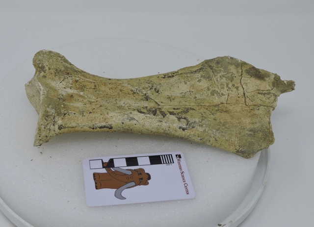 This week we continue our documentation of Pleistocene fossils from the Harveston section of Murrieta, California, with a horse scapula.This specimen is a partial right scapula, shown above in medial view. It's missing a small part of the dorsal edge, on the right in the photo. The part of the scapula is cartilaginous in young animals, and actually only ossifies fairly late in ontogeny, so it's not unusual to be missing this area.
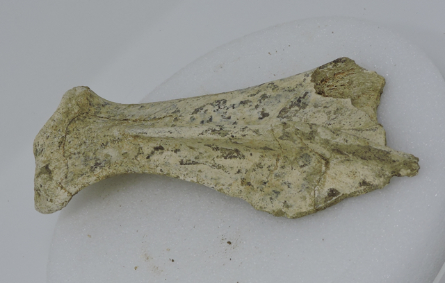 The lateral view is shown above, with anterior at the bottom (this image is missing the scale bar because these are part of a set used to produce a photogrammetric model). The surface on the left is the edge of the glenoid fossa, the articulation with the head of the humerus (the "shoulder socket"). A bone ridge, called the scapular spine, is pointing straight at the camera and divides the scapula into concave anterior and posterior surfaces. The anterior portion is called the supraspinatus fossa, while the posterior part is the infraspinatus fossa; each serves as the attachment points for muscles of the same names, that are involved in rotating the forelimb.I've uploaded a 3D model of this specimen on Sketchfab, available at
 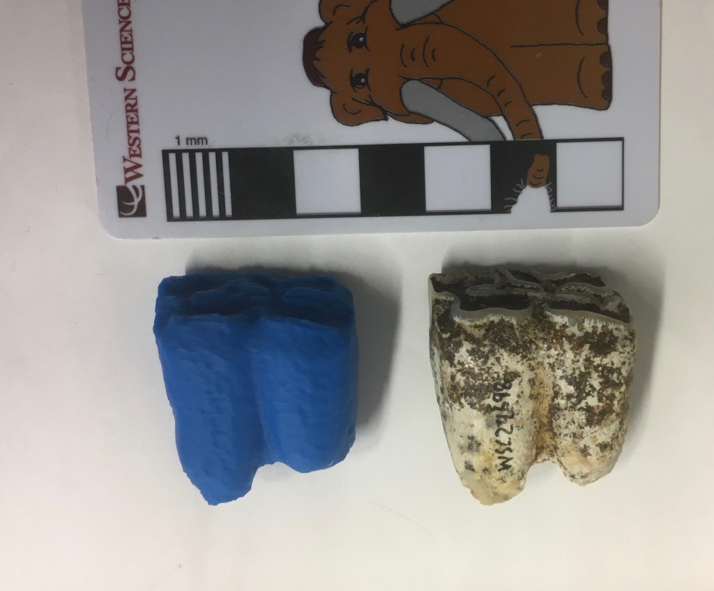 As we continue to work on WSC's collection of Late Pleistocene fossils from Murrieta, it has become clear that, while the collection my be taxonomically diverse, it contains a lot of horse bones!The specimen shown above is a lower right first molar of the horse Equus occidentalis in lateral (labial) view. On the left is an unpainted 3D print of the same specimen. Below is the same specimen in occlusal view:
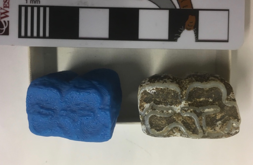 Horses are highly hypsodont, meaning they have very tall, high-crowned teeth. As you can see in the lateral view at the top, we have both the roots and the occlusal surface preserved, showing that we have the whole tooth (it's not broken), yet the tooth is quite short. That's because almost this entire tooth has been worn away, indicating that it came from a very elderly horse.We've obviously scanned this tooth, since we've 3D-printed it. We've also made the scan available on Sketchfab:https://skfb.ly/6xyuR
 We're continuing our efforts to document an describe the fauna from the Harveston neighborhood of Murrieta, a small but diverse collection that appears to be the only Rancholabrean-Age site in Murrieta. The bone shown here is the left lunate of a horse (Equus sp.), with the original bone on the left and a 3D print on the right. The lunate is one of 6 small bones that form the wrist. In horses they are blocky, tightly interlocking bones that can support the stress of holding the weight of a running horse. Unlike primates, which have highly mobile wrists, the horse wrist has essentially no ability to move side-to-side, as is limited to fore-and-aft motion, making it more stable during running.We're planning to scan and print as many of the Harveston bones as possible. The paint job on this print is too dark, so I need to lighten it a bit. O
 We're continuing our review of our collection of Pleistocene fossils from the Harveston neighborhood of Murrieta, in southwestern Riverside County. This is a diverse fauna with a number of different genera, but it appears that in terms of shear numbers of bones this collection is dominated by horses.The specimen shown here is a partial lower jaw of Equus occidentalis, including about 2/3 of the right dentary and a fragment of the left dentary at the mandibular symphysis. At the top is the lateral view, with anterior to the right. Below is the medial view, with anterior to the left:
 And the dorsal view, also with anterior to the left, and showing the occlusal surfaces of the teeth:
 The entire right dentition except for the incisors is preserved, from the 2nd premolar to the 3rd molar. These are all adult teeth, and they are all in wear, so this was an adult horse. In fact, in the lateral view at the top, we can see the unerupted part of the 3rd premolar. There is only a very short crown present above the root. Horses have teeth with extremely tall crowns, so possibly as much as 70-80% of this tooth has worn away. This horse was not just an adult, but was elderly (or, at least its teeth show an amount of wear typically only seen in elderly horses).
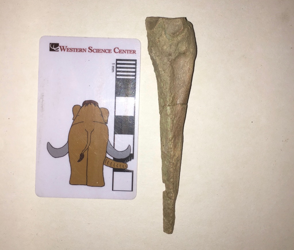
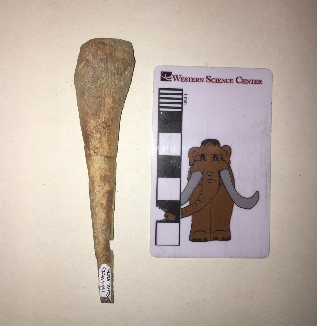 One of the joys of paleontology is that every fossil has a story. Through our understanding of anatomy, geology, ecology, and a host of other field, we can often reveal part of that story, and even a relatively small, nondescript fossil takes on a larger meaning.The small, pointed bone shown above is the right fourth metacarpal from a horse (Equus). The metacarpals are the bones that make up the hand or forefoot. At the proximal end they articulate with the wrist bones (the carpals) and often with the adjacent metacarpals. At the distal end they normally articulate with the first finger bones (the phalanges). In humans, the fourth metacarpal articulates with the ring finger. Our fingers (or digits) are numbered from 1 to 5, with the thumb being Digit I and the pinkie finger being Digit V; thus the ring finger is Digit IV. Toes are numbered the same way, with the big toe as Digit I.Horses famously only have a single hoof on each foot, corresponding to a single finger (on the front feet) or toe (on the back feet). The distal end of the bone shown above (at the bottom of the photo) clearly does not have any kind of articulation for the bones that make up the single finger on the horse's right foot. So what's going on?In horses, the hand bone that supports the animal's weight is the third metacarpal, not the fourth one. The right third metacarpal (probably from the same individual) is shown below:
 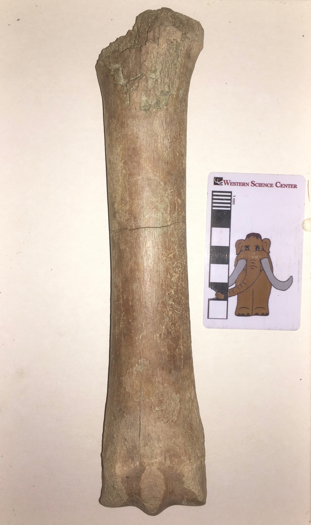 This massive bone is well-suited to holding up a horse's weight. So what about the fourth metacarpal? If it doesn't support a hoof, what does it do?As far as we can tell, pretty much nothing.The fourth metacarpal is a remnant of the horse's evolutionary history. While modern Equus is limited to a single hoof on each foot, supported by Digit III, horses' ancestors and extinct relatives had multiple hooves on each foot. The lateral toes were gradually lost, starting with Digits I and V, and later with Digits II and IV. The key here is gradually. Each digit eventually became small enough that it no longer functioned for walking; there would be a tiny hoof, but it didn't reach the ground. The hoof would eventually be lost, leaving only the metacarpal with no attached finger or toe. After even more time and generations, even the metacarpal is lost. But the whole process takes several million years.In the horse lineage, the first and fifth metacarpals were lost tens of millions of years ago. But the reduction of the second and fourth metacarpals has happened much more recently. Within the last 10 million years horse ancestors still had functional second and fourth toes, even if they were smaller than the central third toe. A few million years ago the second and fourth toes were finally lost altogether, leaving their corresponding metacarpals and metatarsals as the last remaining remnants of these digits. And that's where horses are today. The second and fourth metacarpals (and metatarsals) have lost their toes, and even the articulation to attach to the toes, and they're greatly reduced in size. At the proximal end they still articulate with the wrist bones and the third metacarpal (see the posterior view below). The presence of the fourth metacarpal is evolution caught in the act, a snapshot of a dynamic process that seems static because it happens so slowly. If horses survive a few million more years, the fourth metacarpal will probably be entirely lost, so that there is only a small slice of time (geologically speaking) when such a fossil could be preserved.