

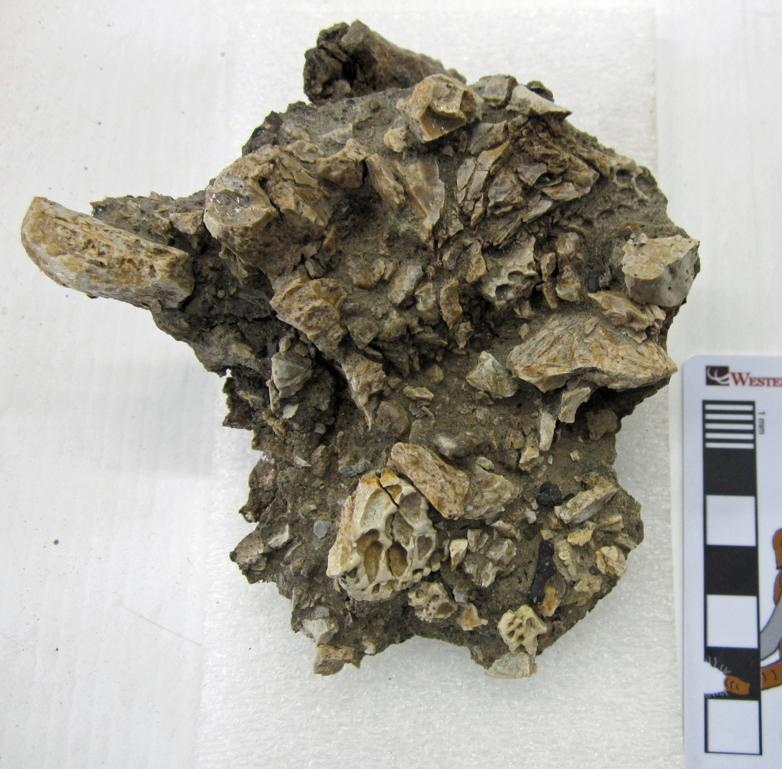
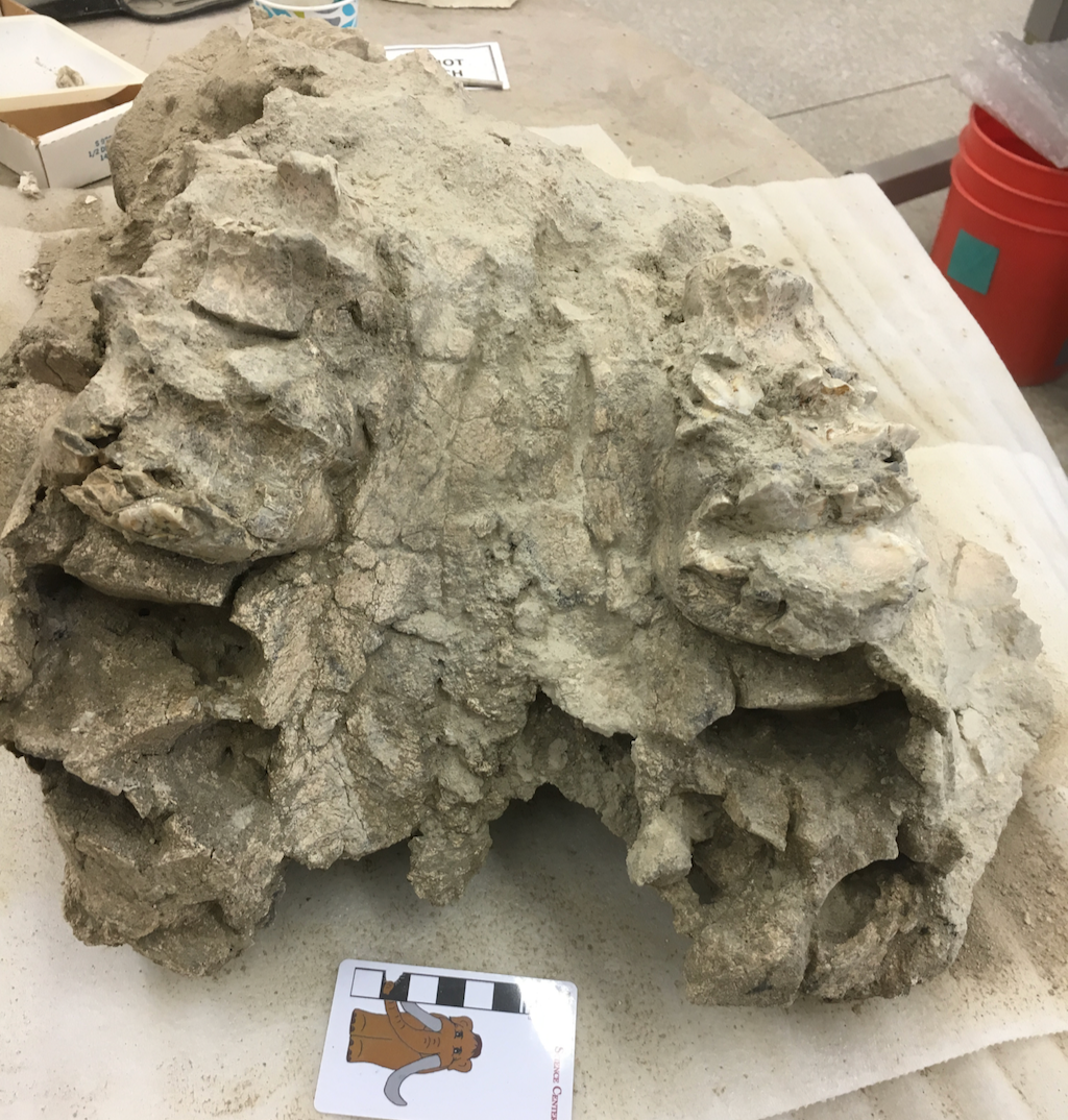
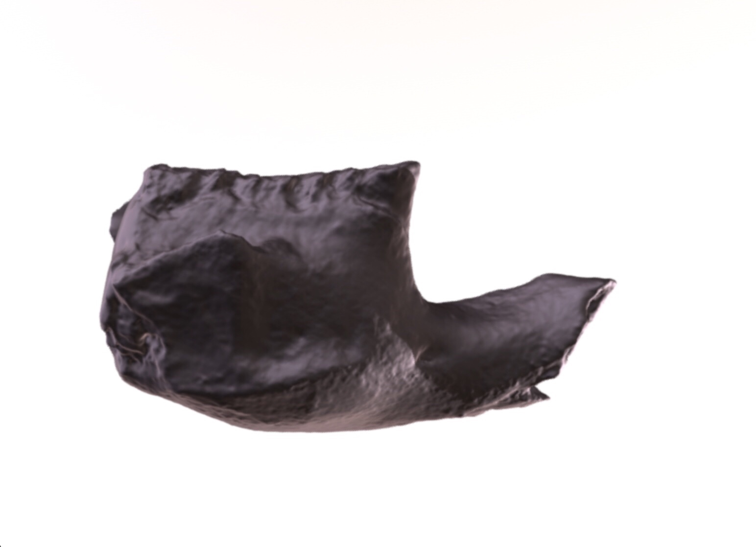
 Giant reptiles abound in our field area in New Mexico. Big dinosaurs like Invictarx, Dynamoterror, and the hadrosaur we're working on now; hefty crocodiles; and even sizable turtles are the most commonly found animals in the 79-million-year-old rocks of the Menefee Formation. Fossils of small creatures are comparatively rare, and skull material for any animal is pretty thin on the ground as well.During the expedition in May with our partners at Zuni Dinosaur Institute for Geosciences, three volunteers from the Southwest Paleontological Society collected a small mass of bone with a distinctive heavily pitted texture that resembles modern crocodilian skulls. In the field, we tentatively identified it as part of a crocodilian skull. We opened up the little plaster jacket here at Western Science Center this week, and confirmed that it is indeed the partial skull of a small crocodilian. The dorsal surface of the skull is badly fragmented, but clearly shows that pitted texture. The ventral side is also highly weathered, but we can see the base of the braincase and the occipital condyle, the ball at the back of the skull that articulates with the first neck vertebra.
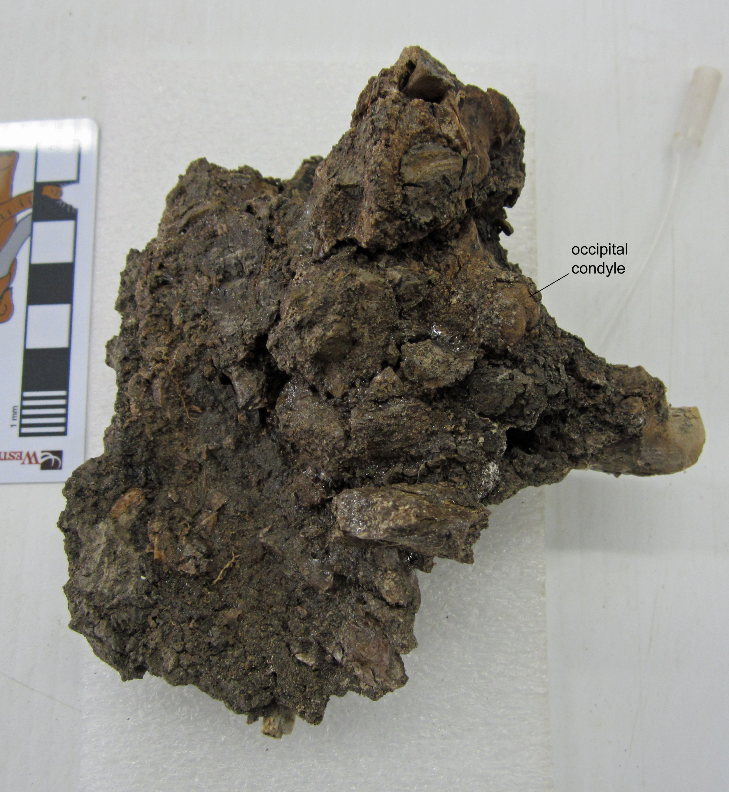 We're just beginning to prep this fossil. Right now, based on the size and skull texture, it might belong to an ancient alligator relative called Brachychampsa. Brachychampsa is known from many Upper Cretaceous formations in western North America, and was first identified in the Menefee Formation of New Mexico by paleontologist Tom Williamson (New Mexico Museum of Natural History and Science) in 1996. It's a small animal, just about six feet long, with a broad, blunt snout, as you can see on this mounted skeleton that I photographed at the Natural History Museum of Utah in Salt Lake City. We'll have much more to say about our little skull and the other Menefee crocodilians in the not too distant future.
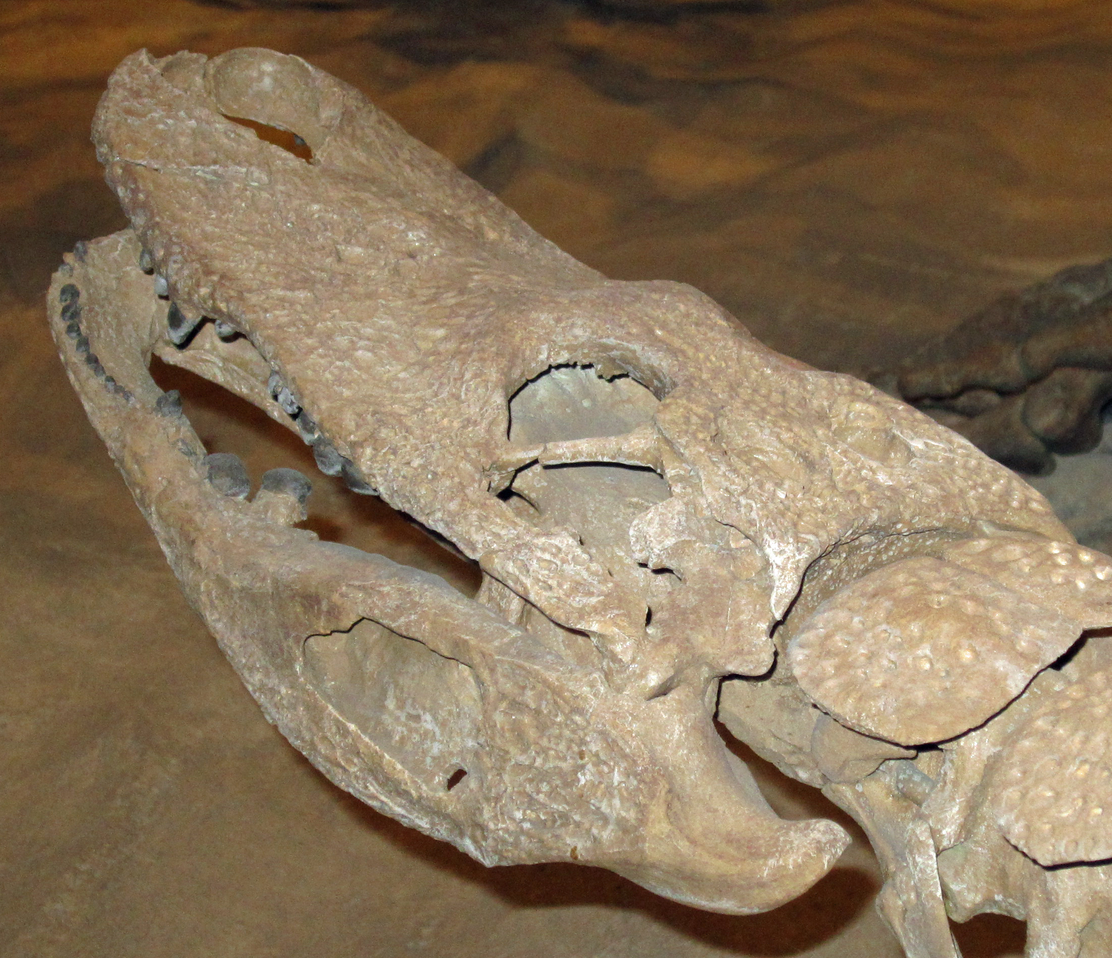 Post by Curator Dr. Andrew McDonald
Fossil Friday - community on a half-shell
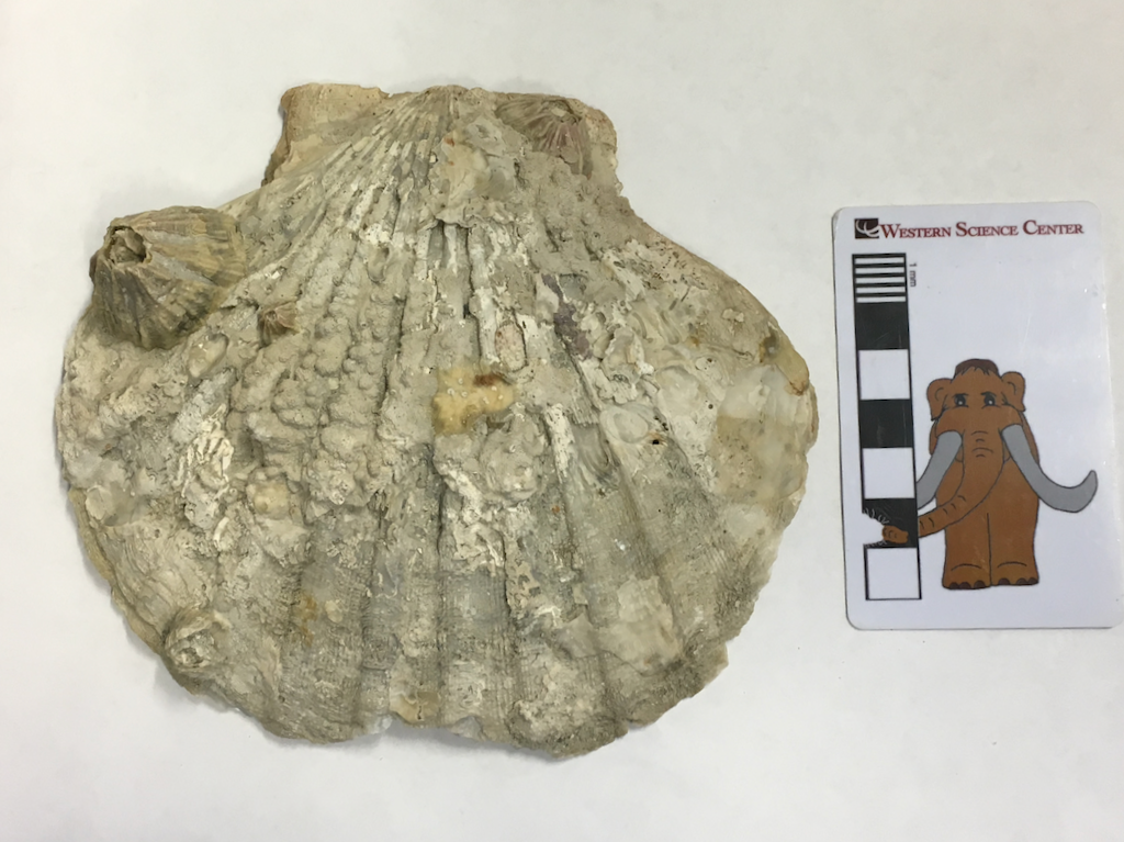 Life as a suspension feeder is a mixed blessing. Most suspension feeders are sessile, staying in one place for practically their entire life. They feed by extracting food particles from water, using a bewildering variety of suction tubes, mucus nets, feathery appendages, and various other anatomical variations. Almost every phylum of animals and many protists include examples of suspension feeders. It's a lifestyle that could be regarded almost as idyllic, as you sway back and forth in the current, waiting on food to blunder into your mouth (for those suspension feeders that actually have mouths; not all do). But it can be treacherous, too. Those same currents that bring you your food can also rip you from your anchor point, carrying you off, where you may ironically end up as food for some other suspension feeder. Attaching to a solid, stable substrate is critical to survival for a suspension feeder, and in some environments there is a lot of competition for good spots.Above is a shell of the extinct scallop Chesapecten madisonius, a species that was abundant in warm-temperate waters of the western Atlantic during the Pliocene. This example came from a quarry near Chuckatuck, Virginia, from the 3-million-year-old Yorktown Formation. Scallops are suspension feeders, although they are capable of moving around. When they died, their large (up to the size of a dinner plate!) calcium carbonate shells would end up lying on the seafloor.At this particular location, the sediment is almost all sand, with huge numbers of broken seashells. Both the sediment and types and preservation of the shells indicate that this was an offshore shoaling area where waves were breaking in shallow water. With the constant water flow this was a great environment for suspension feeders, but the high energy and unconsolidated sandy bottom mean that hard substrates for attachment were at a premium. A large mineralized seashell sitting on the seafloor was too good to pass up, and a whole community of suspension feeders took advantage and settled on the shell.Some of these organisms are gone, but left traces behind indicating their presence. There are several pits at various places on the shell:
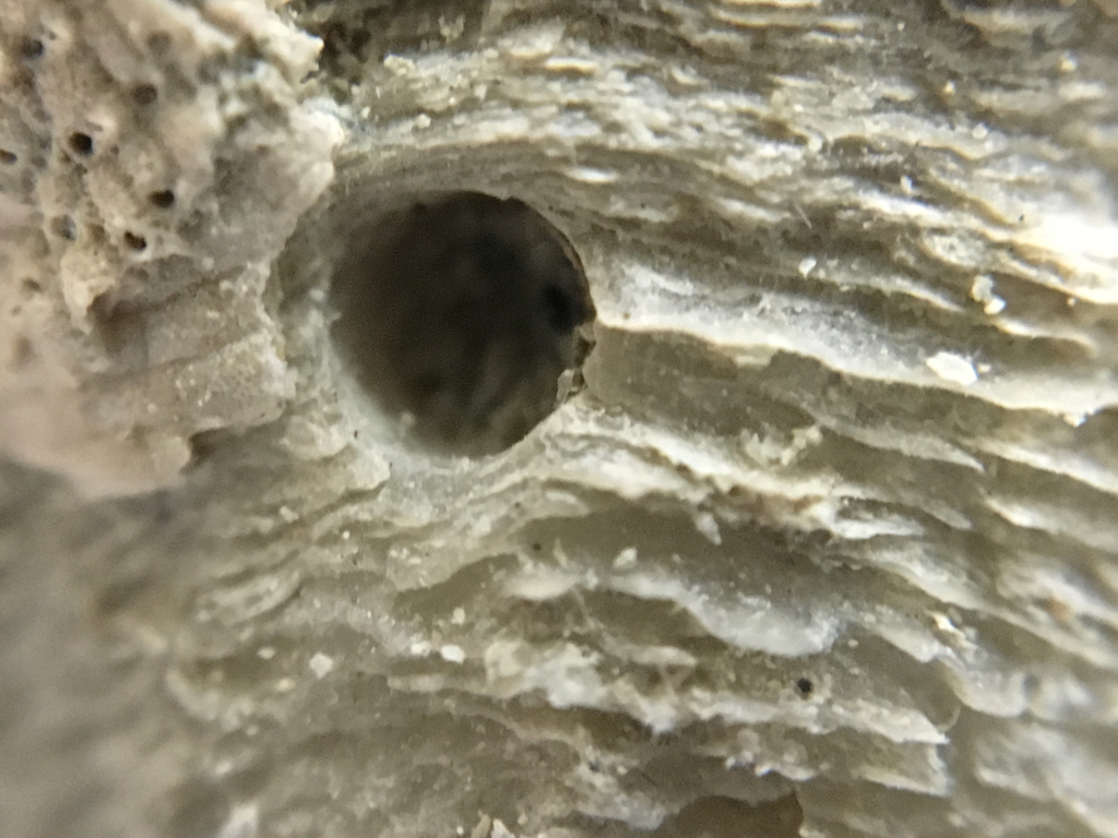
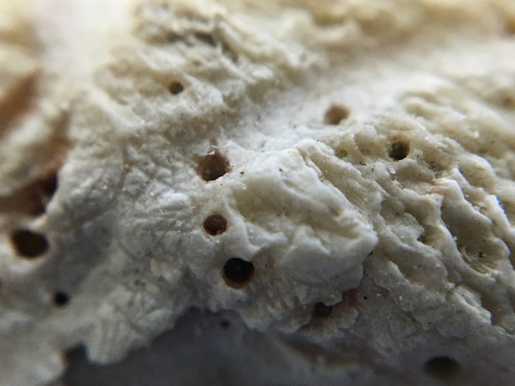 A few of these were probably made by predatory snails that attack other mollusks by drilling into their shells. Others were likely formed by cloned sponges, which chemically dissolve part of the target shell to provide living space.In other areas, parts of the shell have been broadly removed, leaving shallow sculpted depressions:
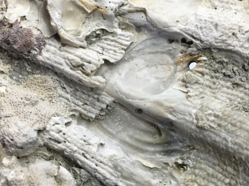 These were likely produced by another mollusk, possibly slipper snails such as Crepidula or boring clams from the family that includes shipworms (which are actually clams). Some were definitely clams of some sort, because one of the shells is still there (although I've not yet been able to identify the species):
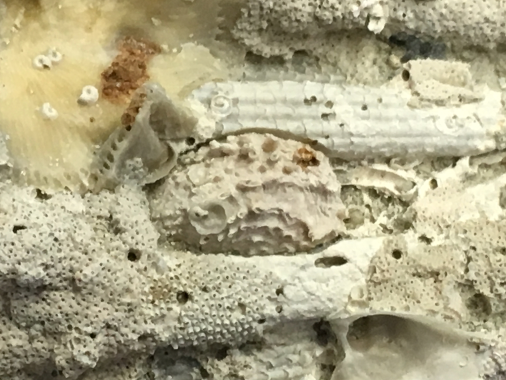 In many cases the colonizing organisms' shells are still present. The most obvious are these volcano-shaped structures:
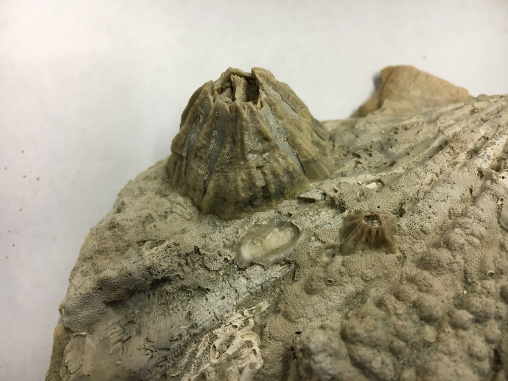 These are barnacles, sessile suspension-feeding crustaceans that are distantly related to shrimp (as a nod to the evolutionary imagination of suspension feeders, they collect food by waving their hairy legs around in the water). Sitting between two barnacles is (I think) an attachment point for an oyster:
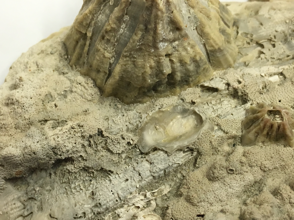 Nearby are small carbonate tubes. I believe these are tubes from polychaete worms (there are also clams that build carbonate tubes, but I'm pretty sure these are from worms):
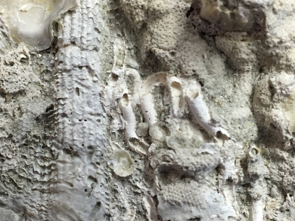 In terms of coverage area, the most extensive colonizers on this shell are bryozoans, or moss animals. These are colonial animals that are placed in their own phylum. Here's one of the larger colonies on this shell:
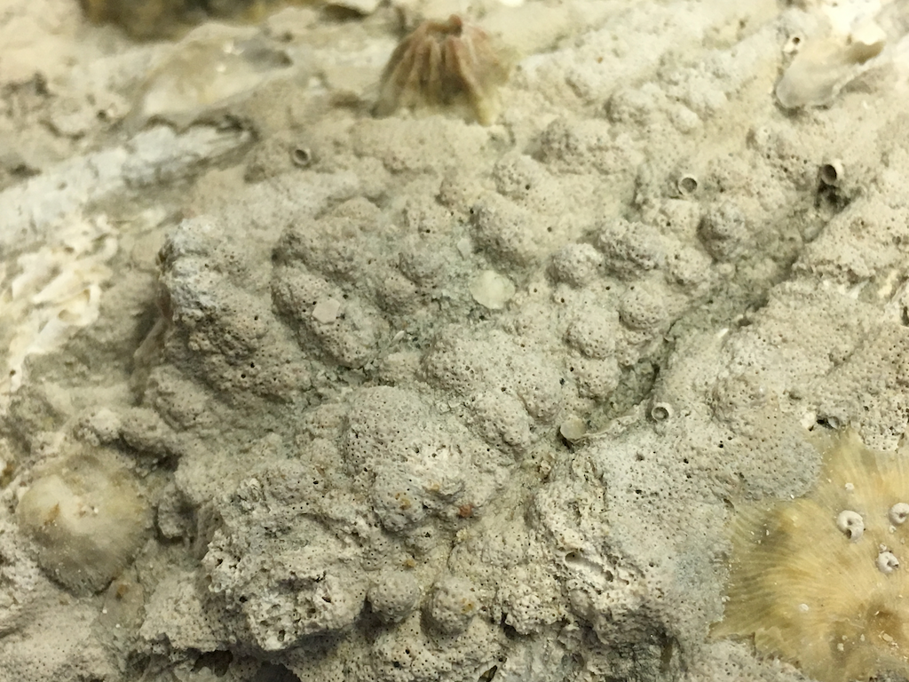 Zooming in close, we can see the individual animals ("zooids") in the colony; each hole contained a zooid:
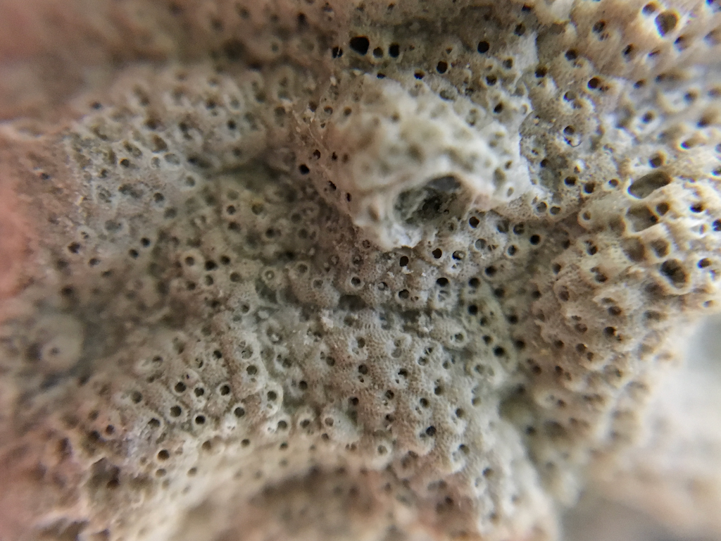 It seems that this shell was colonized by at least two species of bryozoans, because there's another area where the zooids look rather different:
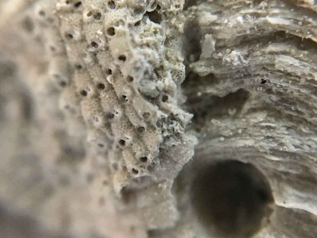 Finally, there's one I'm really uncertain about. There's one patch (the yellowish area at the center of the shell in the top image) where a barnacle attached itself on top of the bryozoan colony, and then fell off (the yellow area is the base of the barnacle). But, on top of that, there are a handful of tiny spiral shells, the largest of which is just under 1 mm in diameter:
 I'm not completely sure of my identification here, but I think these are benthic foraminifera. Foraminifera are amoeba-like, single-celled protists that secrete a calcium carbonate shell (in most species). There are perhaps a dozen of these visible, in addition to places where they were clearly present at one time and later removed.So, assuming all my identifications are correct, and counting everything up, this single Chesapecten shell supported:
- A sponge
- At least a half-dozen bivalves, from at least two species
- At least 10 barnacles
- At least a dozen polychaete worms
- At least two bryozoan species each with several hundred individuals
- At least a dozen foraminifera
Not bad for a single seashell!
Fossil Friday - dinosaur jacket
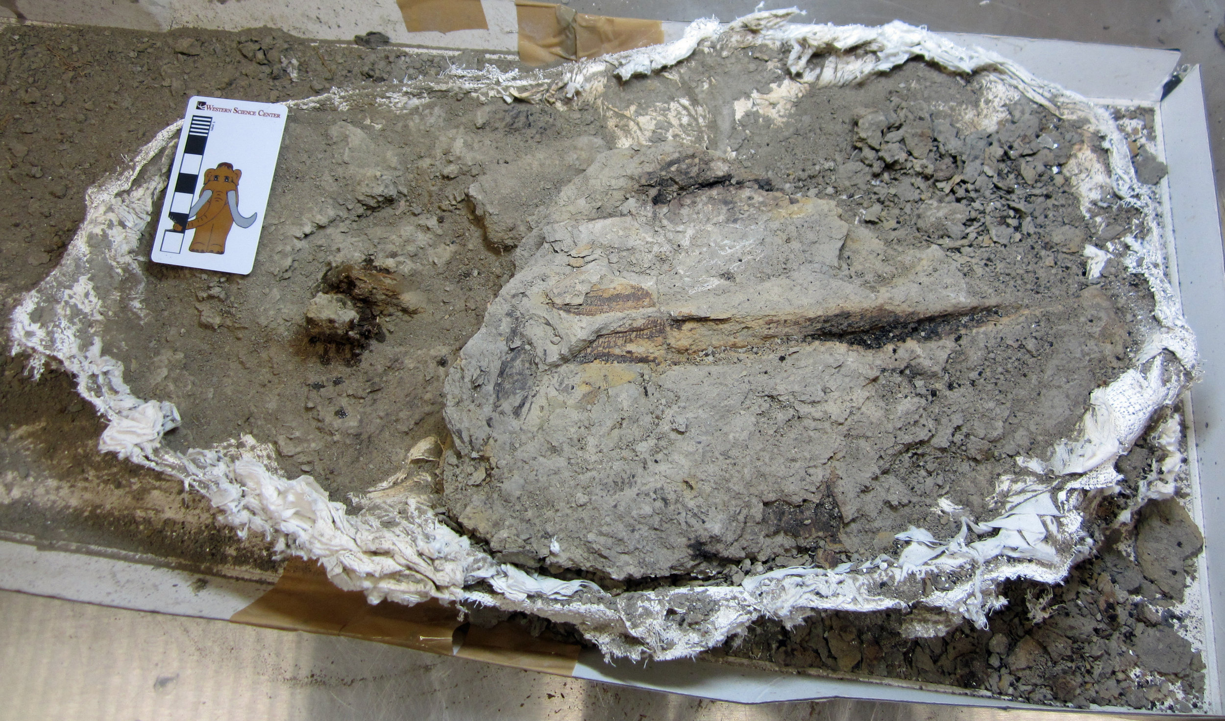 The fossil prep lab at Western Science Center is abuzz these days (literally, when the air scribes are blasting away). WSC staff and volunteers are working their way through the half-ton of fossils we brought back to the museum last summer from the Upper Cretaceous Menefee Formation of New Mexico.Much of our recent prep effort has focused on the hadrosaur skeleton I posted about back in September and October. We're now moving on to some of the smaller items we brought back.One of the new sites we documented and collected during the 2018 field season is a scatter of at least half a dozen dinosaur bones, discovered by WSC volunteer John DeLeon. Although the bones are highly weathered, we collected them all and got them safely back to the museum. Given their close association at the field site and the absence of any other fossil organisms, these bones probably all belong to the same individual dinosaur. John opened up the largest plaster jacket from the site yesterday. There is a little knob of bone exposed to the left in the image; the rest of the bone is underneath the fossilized tree branch to the right. We don't know what type of dinosaur is represented by these bones, but hopefully the answer will emerge as John continues his work.This specimen and all the Menefee fossils were collected by staff and volunteers from the Western Science Center, Zuni Dinosaur Institute for Geosciences, and Southwest Paleontological Society.Post by Curator Dr. Andrew McDonald
Fossil Friday - Squalodon whitmorei
 Usually we use our Fossil Friday posts to talk about specimens that are housed in the WSC collections. But today I'm going to talk about our newest 3D print, the skull of the primitive toothed whale Squalodon whitmorei.Squalodonts are a family of toothed whales that lived in the Oligocene and the first half of the Miocene. At one time it was thought they might be close to the transition between Eocene archaeocete whales and modern dolphins and other toothed cetaceans. It turns out they were their own specialized branch of toothed whales, members of a once widespread group called the platanistoids that has since been mostly supplanted by the delphinoids. They have a somewhat primitive look largely because of their extremely heterodont teeth, including serrated, triangular back teeth (hence the name Squalodon, which literally means "shark tooth"). They were among the last groups of cetaceans (maybe THE last group) to retain these triangular, double-rooted back teeth. They are best known from Europe and eastern North America, but examples have been found all over the world.This specimen holds a special place for me, because it was the first fossil vertebrate I ever had the chance to work on. In 1989 I traveled to the Smithsonian's National Museum of Natural History to talk about possible research topics for my senior thesis at Carleton College. I met paleontologists Clayton Ray, Frank Whitmore, and Dave Bohaska while I was there, and after a day of looking at fossils they suggested that if I wanted to I could come back next summer and work on squalodont whales for my thesis (a group I had never heard of before).The next summer, after securing free housing from a Carleton alumnus, Frank turned over to me several boxes of bone fragments that had been collected by a boy scout troop in Virginia in the 1970s. I spent the next 2 months learning techniques for cleaning and repairing fossils, while Frank tutored my on cetacean anatomy. By the end of the summer I had put together one of the most complete known skeletons of Squalodon, the largest example of the genus ever found:
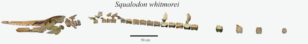
 After successfully defending my undergraduate thesis, I enrolled in graduate school at LSU, and continued my work on squalodonts. This skeleton, along with an additional skull discovered in Virginia in the 1990's, became the main subjects in Chapter 4 of my doctoral dissertation.After completing my doctorate I accepted a position at VMNH, and spent some of my time finishing up projects I had started in graduate school. Finally, in 2005 I formally published "A new species of Squalodon (Mammalia, Cetacea) from the Middle Miocene of eastern North America". That paper established the new species Squalodon whitmorei (named in honor of Frank Whitmore), with this specimen as the holotype.As it turns out, in 2019 and 2020 Western Science Center will be opening two exhibits for which Squalodon whitmorei would be a perfect match. WSC is a Smithsonian Affiliate, so I put in a request through the Affiliates Program to get 3D scans of the skeleton. With their help we arranged to have Bernard Means, a Valley of the Mastodons alum and director of the Virtual Curation Lab at Virginia Commonwealth University visit the Smithsonian and begin scanning the skeleton. While there are still more bones to scan, the cranium has been completed and printed (in six parts) on our Lulzbot printers. Last night I finished painting the printed cranium, which is what I'm holding in the image at the top.We'll be using the printed skull in various education programs at WSC until it goes on exhibit next year.
Fossil Friday - ground sloths & therizinosaurs
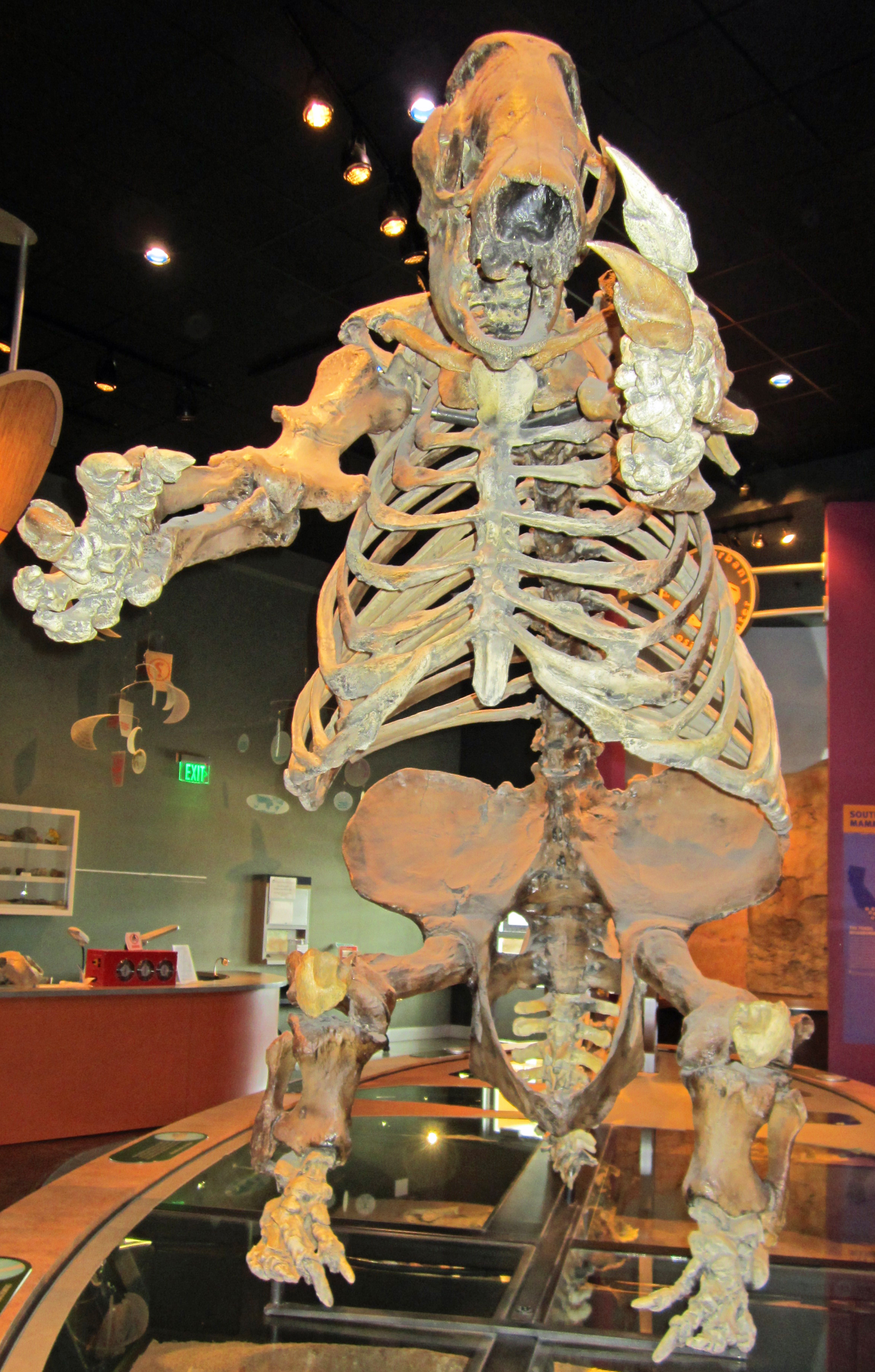 In many ways, dinosaurs and mammals are very different types of animal. And yet, there are some amazing cases of convergent evolution among them. One of the most bizarre is that of giant ground sloths and therizinosaurs. Like the little slow-moving, tree-dwelling sloths of today, giant ground sloths, like this Paramylodon mounted at Western Science Center, were herbivorous. As a group, sloths are related to armadillos and anteaters. Therizinosaurs, like this Nothronychus mounted at the Natural History Museum of Utah in Salt Lake City, are part of Maniraptora, a group of theropod dinosaurs that also includes predators like Velociraptor and our living dinosaurs, the birds. However, therizinosaurs evolved into big feathery herbivores.
 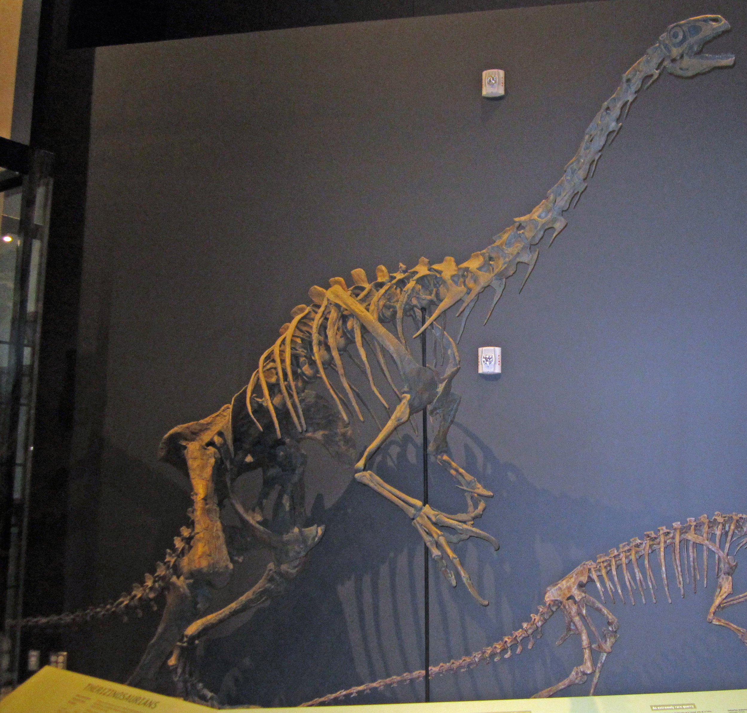 Despite their very different evolutionary origins, giant grounds sloths and therizinosaurs share some remarkable similarities: long arms ending in huge claws, wide bodies to accommodate a long digestive tract for processing plant matter, short robust legs, and short tails. Both groups evolved into large-bodied bipedal plant-eaters. Nothronychus was named in 2001 by my colleagues Jim Kirkland (Utah Geological Survey) and Doug Wolfe (Zuni Dinosaur Institute for Geosciences) - the name means "sloth claw".Another similarity is that both giant ground sloths and therizinosaurs represent migrations into North America from other places. Giant ground sloths evolved in South America and reached North America around nine million years ago. Until the discovery of 90-million-year-old Nothronychus in New Mexico, therizinosaurs were known only from fossils found in Asia. Nothronychus is closely related to the large Asian therizinosaurs and probably represents a migration event into western North America.Post by Curator Dr. Andrew McDonald
Fossil Friday - creodont skull
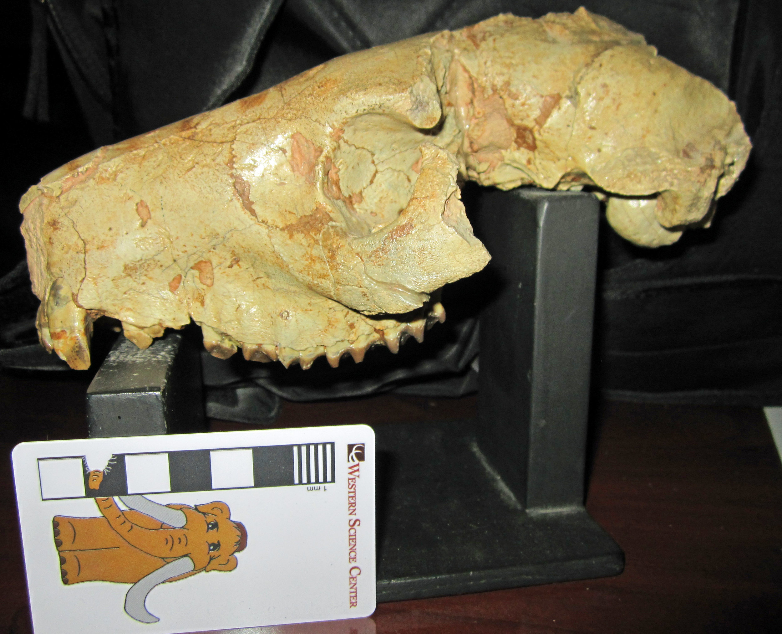 Most of the extinct mammals in the Western Science Center's collection are Pleistocene Ice Age creatures, such as mastodons, Columbian mammoths, bison, camels, horses, and giant ground sloths, dating to between 50,000 and 14,000 years ago. Today's Fossil Friday is a much older extinct mammal, dating back to around 30 million years ago, a slice of geological time known as the Oligocene. This is the skull of an oreodont, a totally extinct group of even-toed ungulates (Artiodactyla) related to camels.This particular skull was discovered in 1963 in South Dakota by renowned local fossil hunter Harley Garbani and donated to the Western Science Center by his wife, Mary. Oreodonts were extremely abundant and diverse plant-eaters in the Oligocene of North America. This skull belongs to an oreodont called Merycoidodon, which was extremely common in South Dakota at this time.
Fossil Friday - juvenile hadrosaur jaw
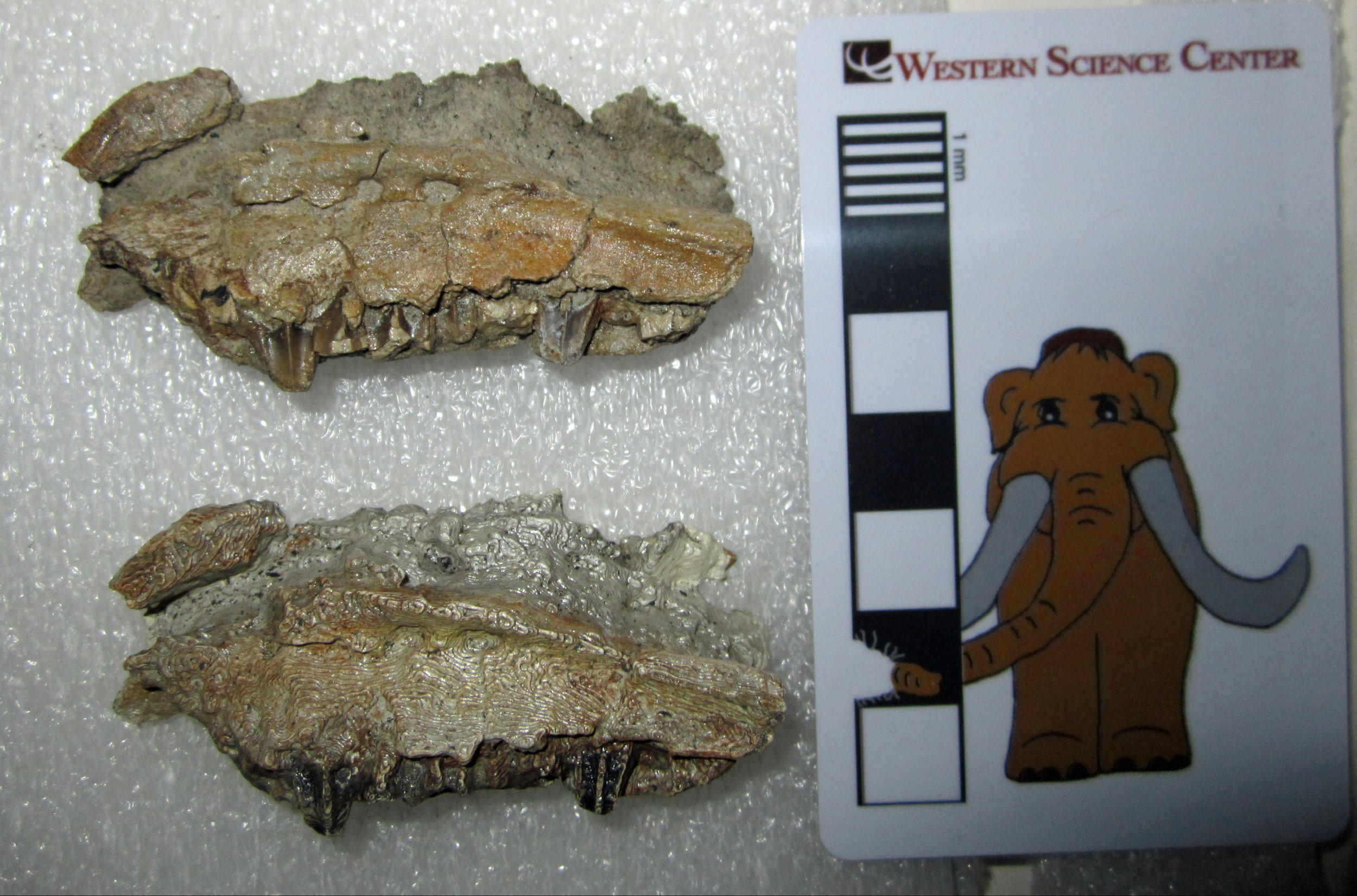 Last week, five staff members from the Western Science Center, including yours truly, traveled to Albuquerque to attend the 2018 Society of Vertebrate Paleontology conference. We presented six posters on the research, outreach, and educational activities taking place at the museum. Three of the posters dealt with fossils discovered in the Menefee Formation of New Mexico, including the new dinosaurs Invictarx (https://valleyofthemastodon.wordpress.com/2018/08/24/fossil-friday-invictarx-zephyri/) and Dynamoterror (https://valleyofthemastodon.wordpress.com/2018/10/12/fossil-friday-dynamoterror-dynastes/). We presented this research with our colleagues from Zuni Dinosaur Institute for Geosciences and Southwest Paleontological Society.This beautiful little bone was the subject of a poster led by Kara Kelley, who has been participating in Menefee expeditions since 2012 and is now an undergraduate at the University of Arizona - Tucson. It's the left maxilla (upper jaw bone) from a very young, very small hadrosaur, which Kara discovered in 2016. Bones of baby dinosaurs are often rare, and this remains the only such bone we have collected in the Menefee Formation so far. In the image, the outer surface of the bone is shown, with a row of tiny teeth along the bottom. The animal's snout would be towards the left of the image, and the back of the skull towards the right. We can't yet tell exactly what kind of hadrosaur this little maxilla belongs to, but it does provide information on the anatomy of a young hadrosaur that lived 80 million years ago.And lest you think that we have two tiny hadrosaur maxillae, the bottom one is actually a painted 3D-print that we created here at Western Science Center through photogrammetry. Now we have a replica of the maxilla that we can take to events and conferences to show people and highlight just one of the many research projects unfolding here at the museum.Andrew McDonald
Fossil Friday - Dynamoterror dynastes
Welcome Dynamoterror dynastes!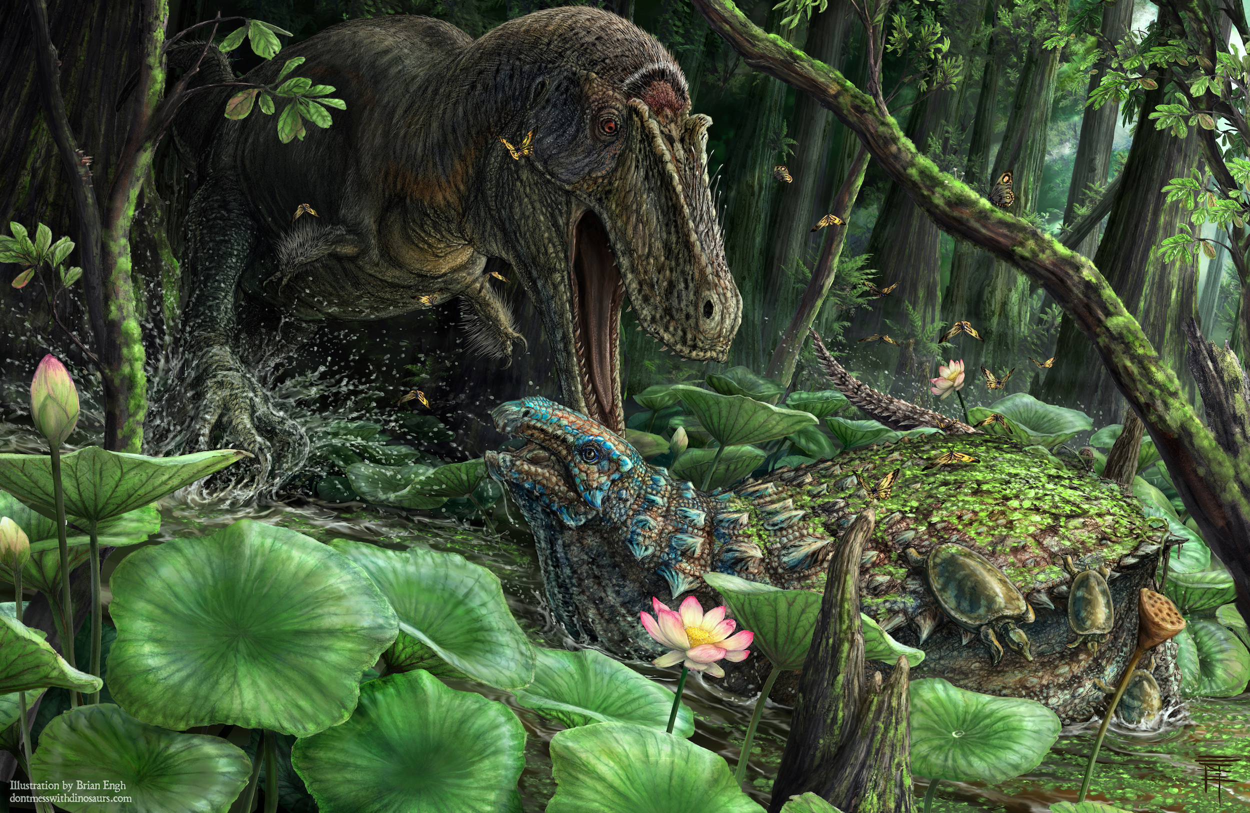 This is another very special Fossil Friday for Western Science Center and our colleagues at Zuni Dinosaur Institute for Geosciences and Southwest Paleontological Society. Hot on the heels of our new armored herbivorous dinosaur, Invictarx zephyri comes this new tyrannosaur. Like Invictarx, Dynamoterror lived 80 million years ago in what is now New Mexico and is published in the online open-access journal PeerJ. Dynamoterror is known from an incomplete skeleton that includes bones from the skull, hand, foot, and many other fragments.
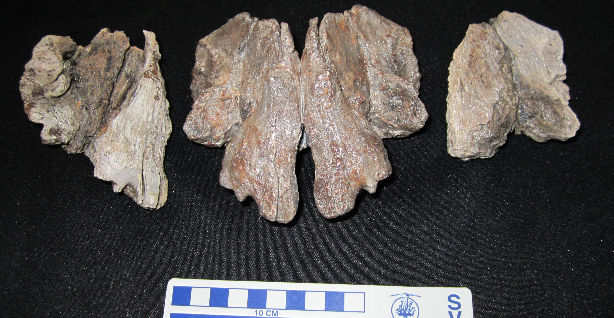 The first image shows the two frontal bones from the top of the skull of Dynamoterror (left and right) and the 3D-print of the composite frontals created at Western Science Center (center). We made the 3D-print by laser-scanning the actual fossils, building 3D digital models of them, and then mirroring and combining the models based upon overlapping anatomical features preserved on both frontals. This 3D-print helped us refine our interpretation of the anatomy in this region of the skull and is a nifty item to share at public events!Dynamoterror lived at the same time and place as Invictarx and the hadrosaur I have posted for Fossil Friday the last three weeks, and probably was quite a threat to them. Paleoartist Brian Engh has depicted a less than friendly encounter between Dynamoterror and Invictarx in a spectacular piece of artwork.Post by Curator Dr. Andrew T. McDonald.
Fossil Friday - hadrosaur update #2
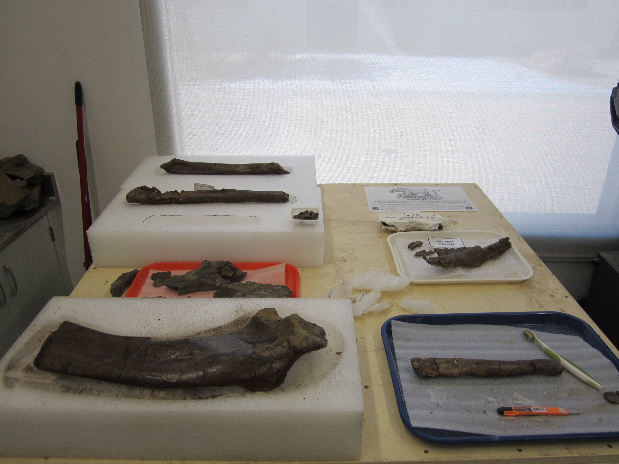 For the last two weeks, I've been giving you updates on the preparation of a big block of rock packed full of hadrosaur bones from New Mexico.All seven bones have now been freed and Western Science Center staff and volunteers are putting the finishing touches on them - removing the last bits of rock, repairing breaks, and reinforcing cracks.
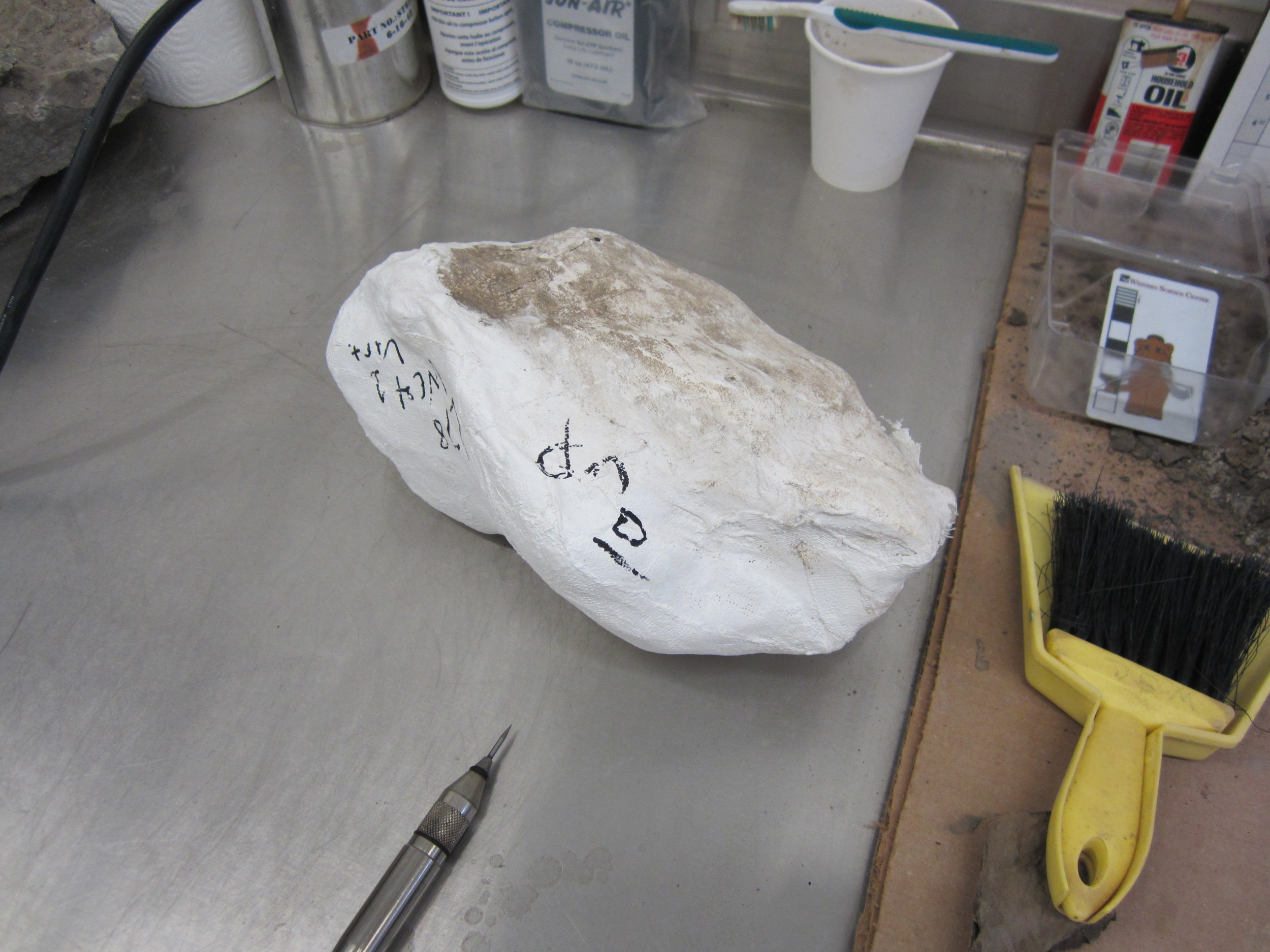 Volunteer Joe Reavis, who worked extremely diligently on the big block for over a month, is now moving on to some smaller jackets from the same quarry, starting with the small jacket pictured here. There are two more jackets twice the size of this one! What parts of this 80-million-year-old herbivorous dinosaur do they contain? I'll continue to post updates as more of this remarkable animal is freed from the mudstone. Post by Curator Dr. Andrew T. McDonald
Fossil Friday - hadrosaur jacket update
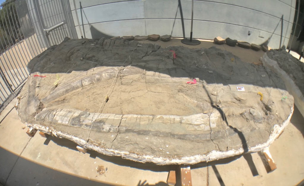
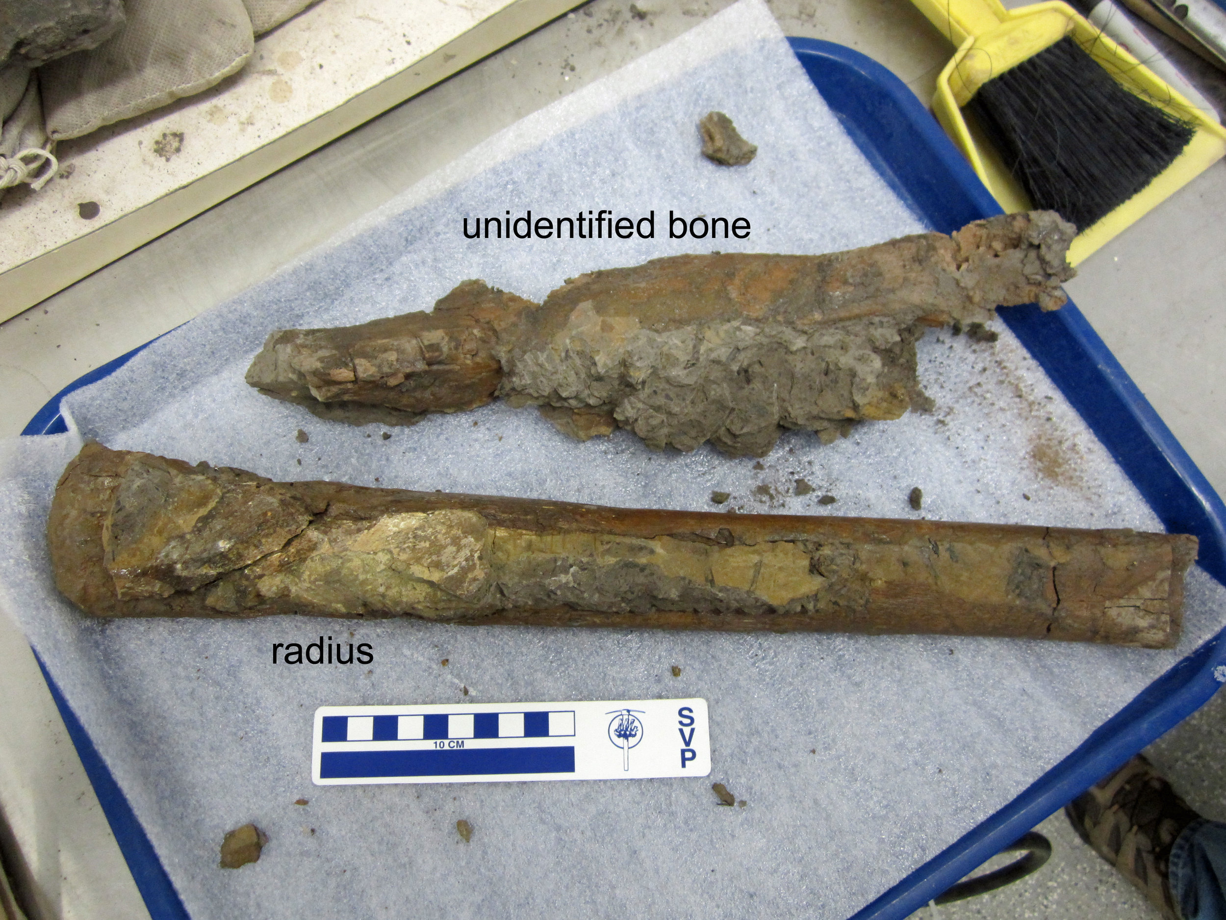 Last week for Fossil Friday, we posted about a big plaster jacket packed full of 80-million-year-old hadrosaur bones, which we brought back to the Western Science Center from New Mexico in June.Volunteer Joe Reavis has been working very diligently on removing the rock from the large but quite fragile bones. Every week brings more progress and greater understanding of which parts of the animal are actually present.Since last Friday, Joe has separated the two forearm bones from the scapula. It was all hands on deck earlier this week as we carefully lifted the two forearm bones away from the jacket, leaving the scapula completely intact. Last week, I identified the two long forearm bones as the left and right radii (https://valleyofthemastodon.wordpress.com/2018/09/21/fossil-friday-hadrosaur-bones/#more-1697). However, one of them is not a radius after all, but is instead an ulna, the other long bone in the forearm.
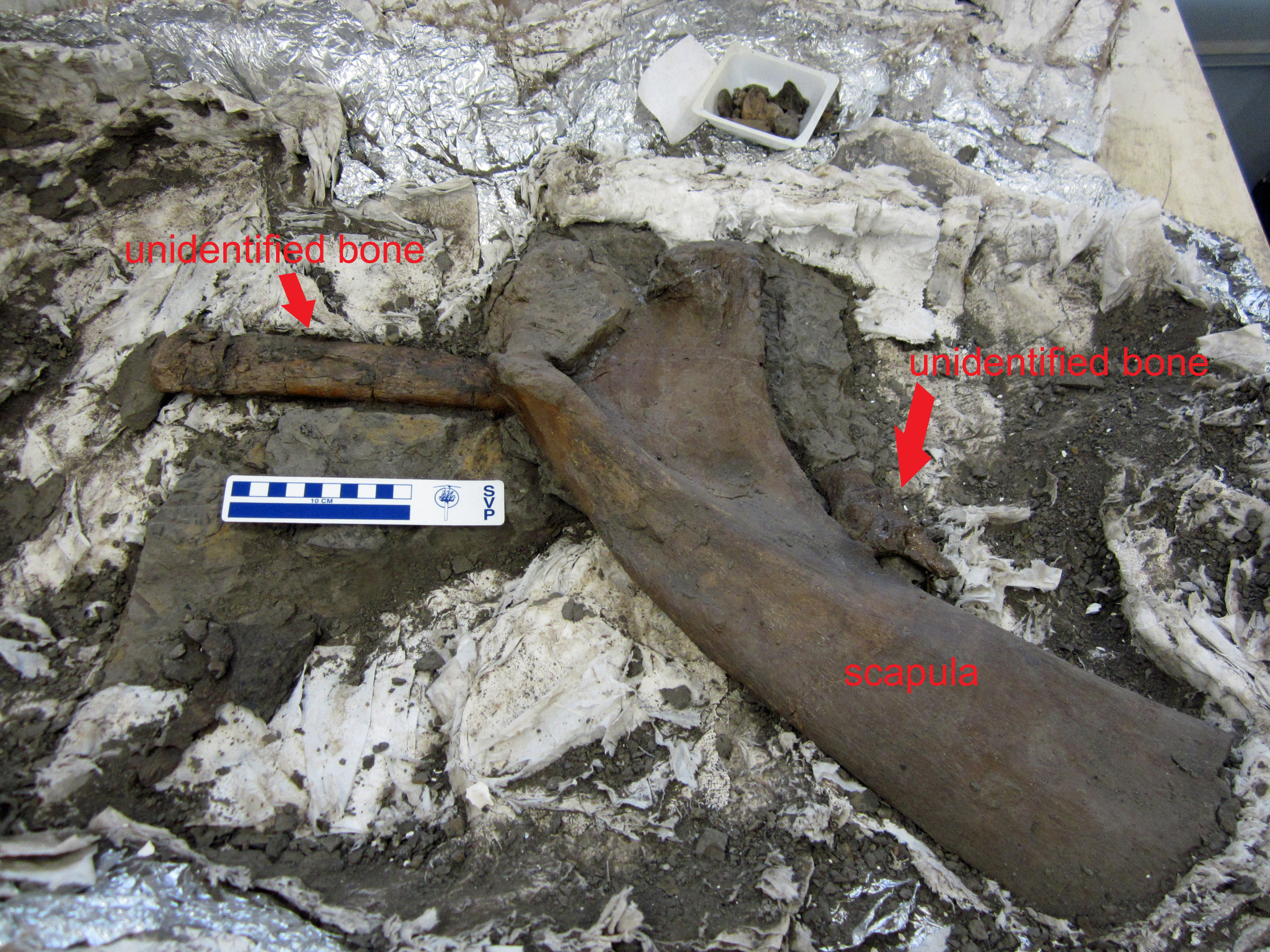 Finding fossils in the field is not the end-all and be-all of discovery. Instead, it's only the first step. As we continue to clean the bones of this hadrosaur skeleton, we will refine the identities of the preserved bones and eventually start to investigate what kind of hadrosaur we have sitting in our lab.
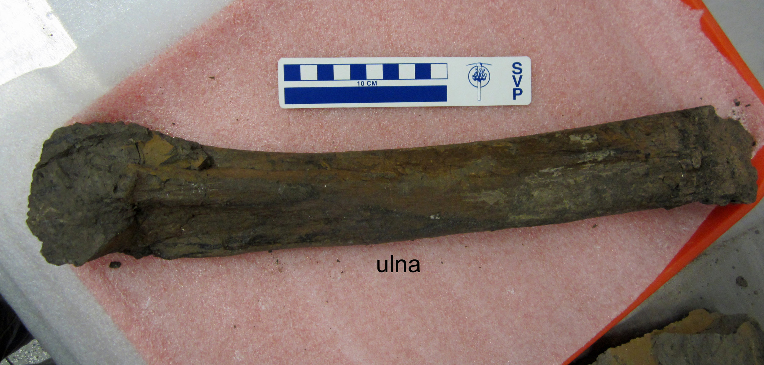 Post by Curator Dr. Andrew McDonald.
Fossil Friday - hadrosaur bones
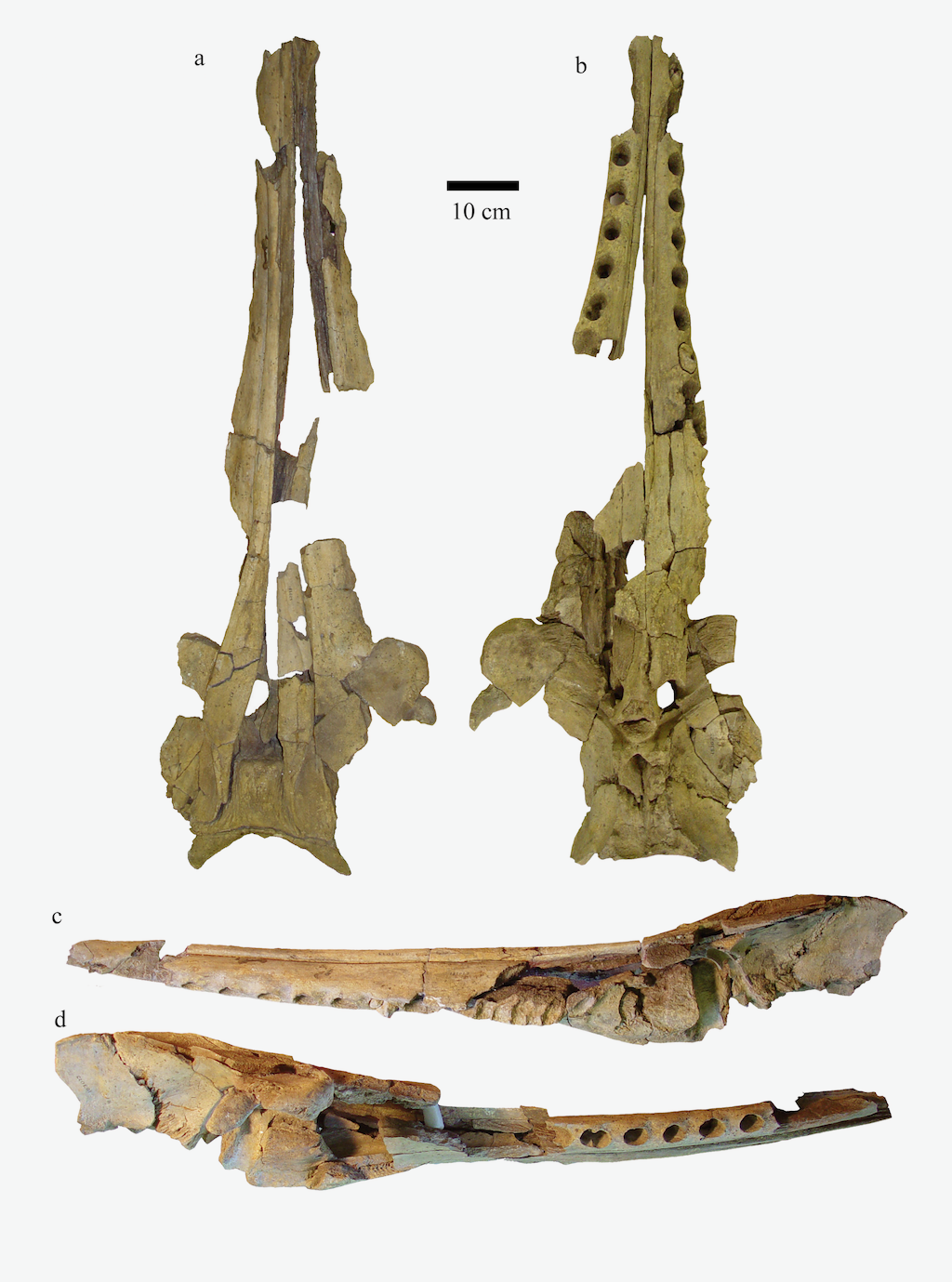
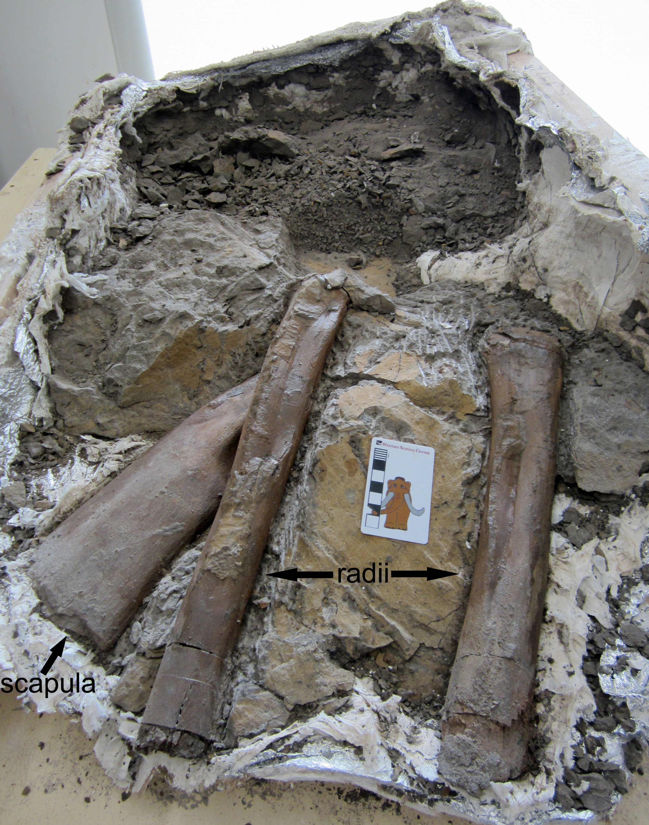 Being wrong is part of the scientific process. Scientists make new discoveries every day, and sometimes prior ideas are supported and sometimes they are refuted as we learn more.Today's Fossil Friday is a perfect example of this, as well as being a really exciting fossil! In June, my colleagues and I returned to the Western Science Center after three weeks of field work in New Mexico. Among the half-ton of 80-million-year-old fossils we brought back was a large plaster jacket containing what I had identified as several bones of a theropod, some sort of medium-sized meat-eating dinosaur.For the last month, Western Science Center volunteer Joe Reavis has been preparing this big block of rock, carefully revealing the bones within. His work has led to a bit of a surprise - the bones are not those of a meat-eating theropod, but instead a plant-eating hadrosaur! Hadrosaurs are often nick-named the "duck-billed" dinosaurs, because of the wide beaks at the front of their mouths. These herbivores could reach 30 feet or more in length and were extremely common in North America during the last 20 million years of the age of dinosaurs, right up to the mass extinction 66 million years ago that wiped out all the dinosaurs except modern birds.Isolated hadrosaur bones are abundant in the Menefee Formation, the layer of rock we are studying in New Mexico. However, this large jacket contains at least three limb bones in close proximity. So far, Joe has uncovered the right scapula (shoulder blade) and both radii (the radius is a long bone in the forearm). This next image shows the arms of AMNH 5886, a skeleton of the later hadrosaur Edmontosaurus annectens, which I photographed at the American Museum of Natural History in New York. The right scapula and both the left radius and right radius are labelled.
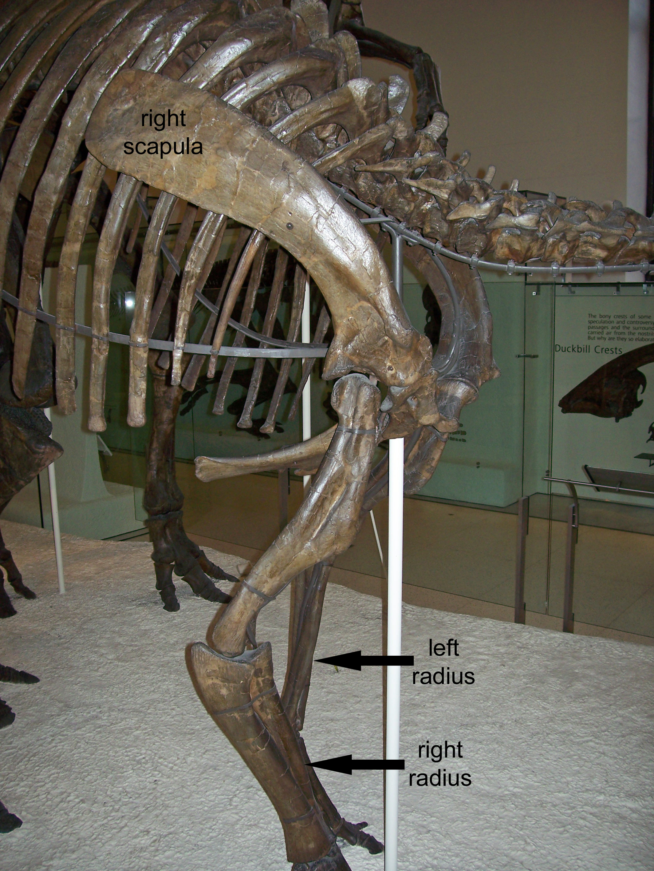 There is still more rock to be removed in the big jacket, and we brought back half a dozen smaller jackets containing other bones from the same site, one of which contains a hadrosaur vertebra. So, we will have much more to say about this remarkable animal in the future. The fossils were collected by staff and volunteers from the Western Science Center, Zuni Dinosaur Institute for Geosciences, and Southwest Paleontological Society.Post by Curator Dr. Andrew T. McDonald
Fossil Friday - camel tooth
 Every day on my way to work, I drive past a farm that among its denizens counts several camels. Apart from the fun of seeing these large, strange mammals, they also serve as a reminder that wild camels once roamed across North America, until their extinction around 12,000 years ago. Although today, wild camels and their relatives are found in Asia, Africa, and South America, in a bit of evolutionary irony, camels actually originated in North America and spread around the world from there. The first camels appeared around 35 million years ago during the Eocene Epoch, in the form of small fast-running herbivores like Poebrotherium. By the Miocene Epoch, camels had diversified and grown larger. This image shows one of the upper left cheek teeth of a camel that lived here in California between 18 and 14 million years ago. This tooth was collected by the Western Science Center earlier this year at a fossil site in San Bernardino National Forest. It might belong to an extinct camel called Aepycamelus, which was widespread in North America during the Miocene. However, we are still studying this tooth to determine exactly what species it represents.
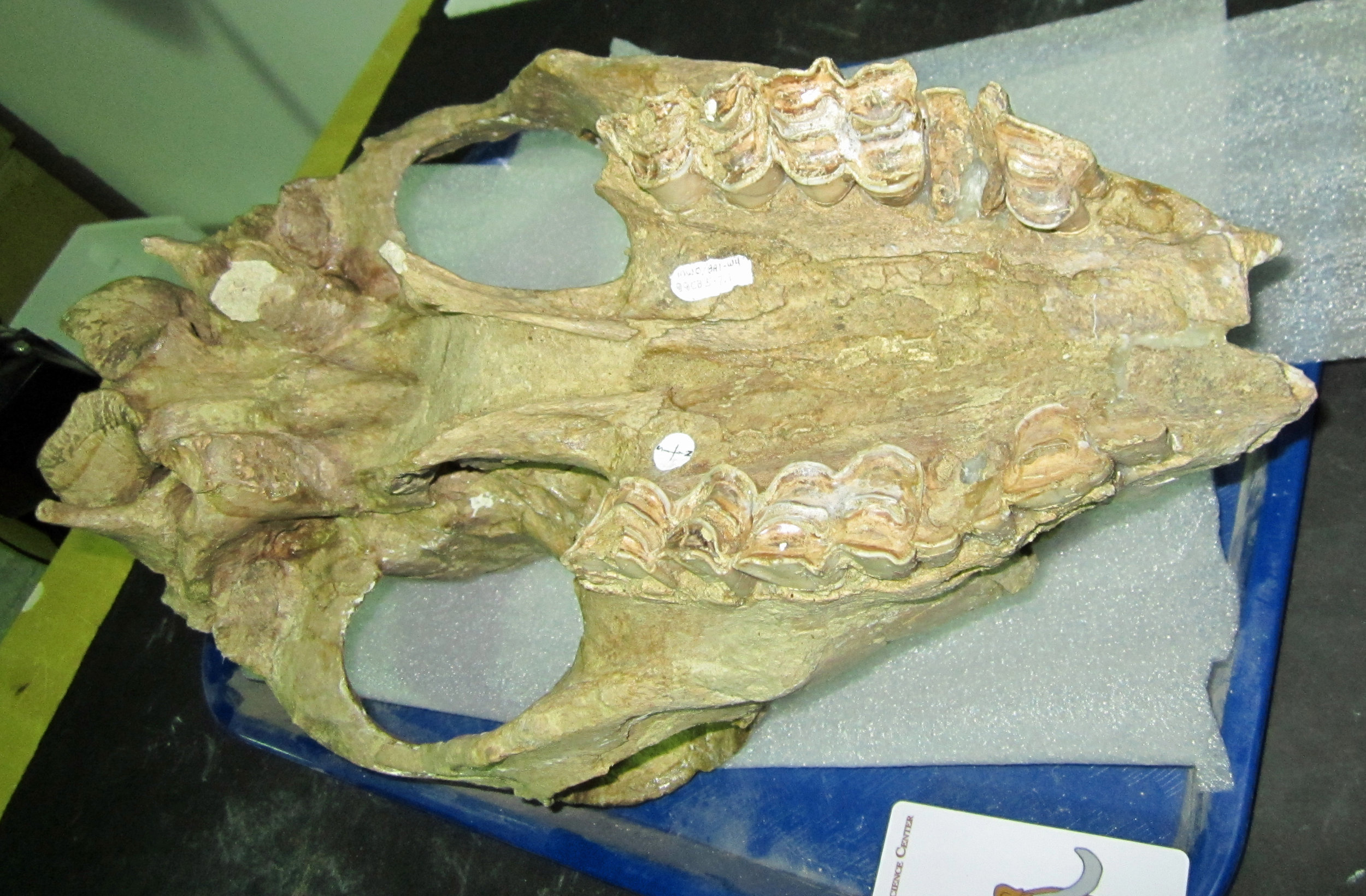 Camels kept going strong in North America up until their extinction at the end of the last Ice Age. One of the last camels in North America was the large species Camelops hesternus, which ranged across western Northern America, including California. This image shows the underside of a Camelops skull discovered at Diamond Valley Lake and which is part of the Western Science Center's collection. The teeth are highly worn, but still bear a close resemblance to the smaller Miocene tooth. Post by WSC Curator Dr. Andrew T. McDonald
Fossil Friday - mastodon skull update
 A few weeks ago I wrote about a mastodon skull we had moved into the lab for additional prep work. Kind of a lot has happened since then.We managed to successfully remove the remaining part of the original field jacket, which had bee on the skull since it was first found in 1997. That enabled us to turn the skull over and start working on the ventral side, where we could get a better view of the teeth.I speculated last week that this skull was almost certainly either a juvenile or a female, based on its small size and small tusk diameter (while the tusks aren't preserved, their sockets are). The determination between juvenile and female depended on whether or not the preserved tooth was the 1st molar (indicating a young animal) or the 2nd molar (indicating a young adult, and therefore probably a female). It turns out everyone was wrong!
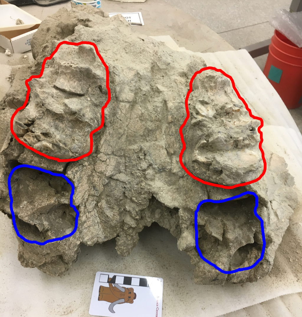 The teeth are outlined in blue above, with the empty tooth sockets outlined in blue. The teeth turned out to be 3rd molars, making the animal much older than I thought! The empty sockets, instead of being for the 4th premolars or 1st molars, were actually for the 2nd molars.The 3rd molars in this case were either unerupted or just starting to erupt; the 2nd molars probably fell out after death. That means this mastodon was probably between 25 and 30 years old when it died. Instead of the pre-teen or teenager I expected, this was a fully mature female mastodon, the first one identified from Diamond Valley Lake.We still have a bit more cleaning to do to the skull. Unfortunately, while there is a substantial amount of bone present behind the palate, it is completely splintered and probably can't be reconstructed. Even so, we'll be getting some good data from this specimen.
Fossil Friday - Invictarx zephyri
 Welcome Invictarx zephyri! This is a very special Fossil Friday for me and everyone here at Western Science Center, and our colleagues at Zuni Dinosaur Institute for Geosciences and Southwest Paleontological Society. The paper naming and describing this new armored plant-eating dinosaur was published today in the online, open-access journal PeerJ. These are the fossils of Invictarx laid out, including vertebrae, parts of the forelimbs, and many osteoderms, the bony armor plates that would have been set in the animal's scaly hide - picture the armored back of an alligator. The image is a composite of three different fossil skeletons found at different spots but in the same rock layer in northwestern New Mexico, and all belonging to Invictarx zephyri.
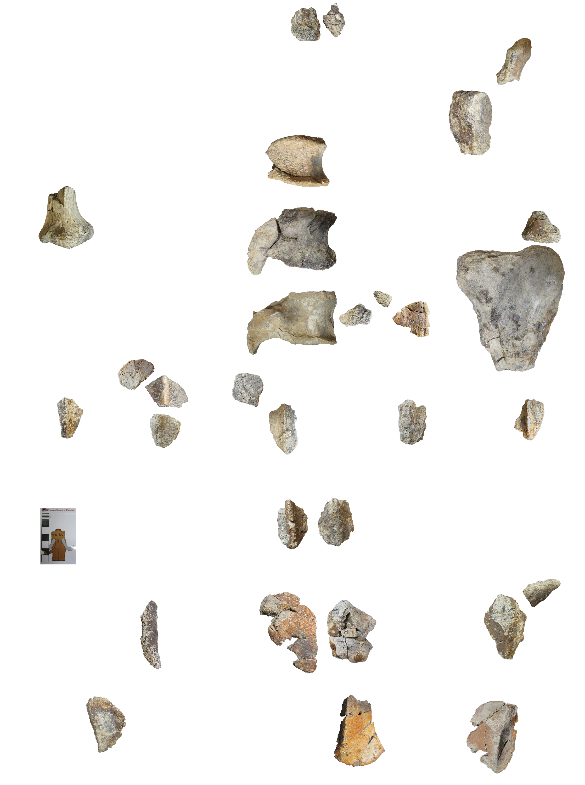 Invictarx lived 80 million years ago, when New Mexico was covered in lush forests, dank swamps, and sluggish rivers, a far cry from the parched badlands in which my colleagues and I found the fossil remains. Invictarx is a nodosaurid, a group of armored dinosaurs that includes many species from North America and Europe. The osteoderms of Invictarx have a unique set of features that set them apart from all other nodosaurids, indicating that we have a new species on our hands, one never found by paleontologists before. Like other nodosaurids, Invictarx sported armor plates across its neck, back, and hips, which probably provided a formidable defense against predators like tyrannosaurs. We collected the first skeleton of Invictarx in 2011, at a site perched atop a high ridge whipped by 40 mile-per-hour winds, which made for a challenging excavation. With all this in mind, my sister and I took out our Latin dictionaries and came up with the name Invictarx zephyri, the "unconquerable fortress of the western wind".
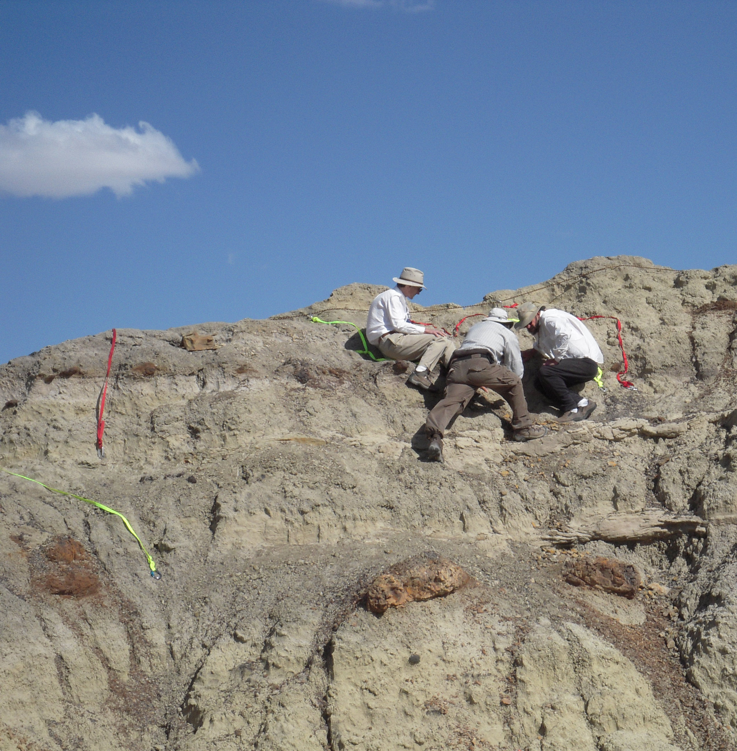 Apart from being a fascinating animal in itself, Invictarx is also the first new species of dinosaur named from the Menefee Formation, the rock layer in which it was discovered. It is the first of many fossil animals and plants from the Menefee that we will describe over the coming years. Of the three specimens, we chose one to serve as the holotype of Invictarx. The holotype of an organism, living or extinct, is the specimen to which the organism's scientific name will always be attached. It serves as the standard by which all future discoveries will be assessed as to whether they represent the same species as the holotype or not. The holotype is accessioned here at the Western Science Center, and is the first holotype in our collections.
  My colleagues and I return to New Mexico every summer to search for dinosaurs and other fossils in the Menefee Formation. We will very probably find additional fossils of Invictarx that will tell us more about its appearance and evolution. Naming it was really just the first step!Post by Curator Dr. Andrew McDonald
Fossil Friday - mastodon skull
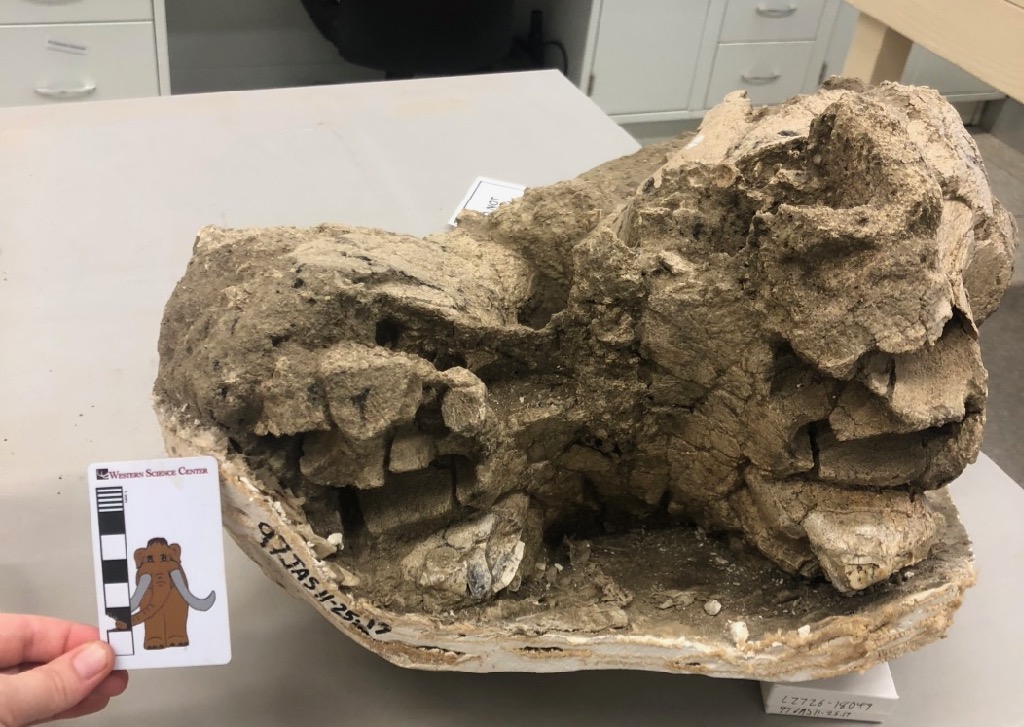 We haven't talked much about mastodons on the blog lately, but that doesn't mean they've been forgotten. We've been doing a lot of work scanning, measuring, and otherwise documenting the mastodons in our collection, and we're doing additional preparation work on some of them. The specimen featured in this week's post was moved into our preparation lab a few days ago for additional work.This may seem at first glance like a nondescript lump of bone, but it's actually a fairly significant amount of a small mastodon skull. This is an unusual view, looking head-on almost straight into the mouth. Below is an annotated version:
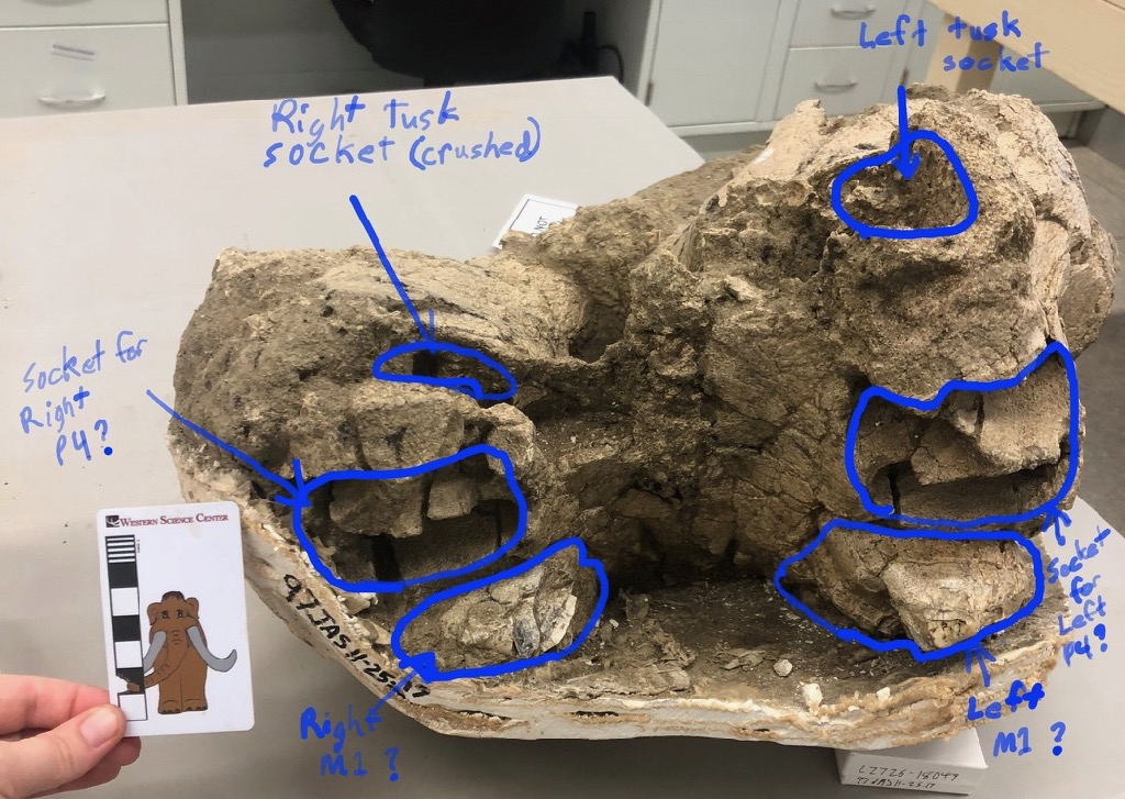 So we actually have nearly the entire palate of this animal, as well as the tusk sockets. Unfortunately, we can't see the teeth clearly because obscured by the plaster jacket, which is why we've moved this into the lab. We're going to try removing the jacket and cleaning the ventral side of the skull. That should enable us to determine if the preserved teeth are in fact the first molars (as suggested in the annotation), or whether they're actually the second molars.Determining the identification of this tooth has broad implications for this specimen. If you noticed the scale bar, you might have realized that this is a very small skull for a mastodon. If the tooth is the 1st molar, that means this mastodon was only about 14-15 years old, a young adolescent. But if it's the 2nd molar, it means that the mastodon was more like 27-28 years old, a full grown adult. To be that small at that age, this would have to be a female (male mastodons are much larger than females).Both female and juvenile mastodons are exceptionally rare from Diamond Valley Lake. In fact, whichever this specimen turns out to be, it will be the only skull from DVL in that category. So, female or juvenile? As soon as we find out, we'll let you know in a future post.
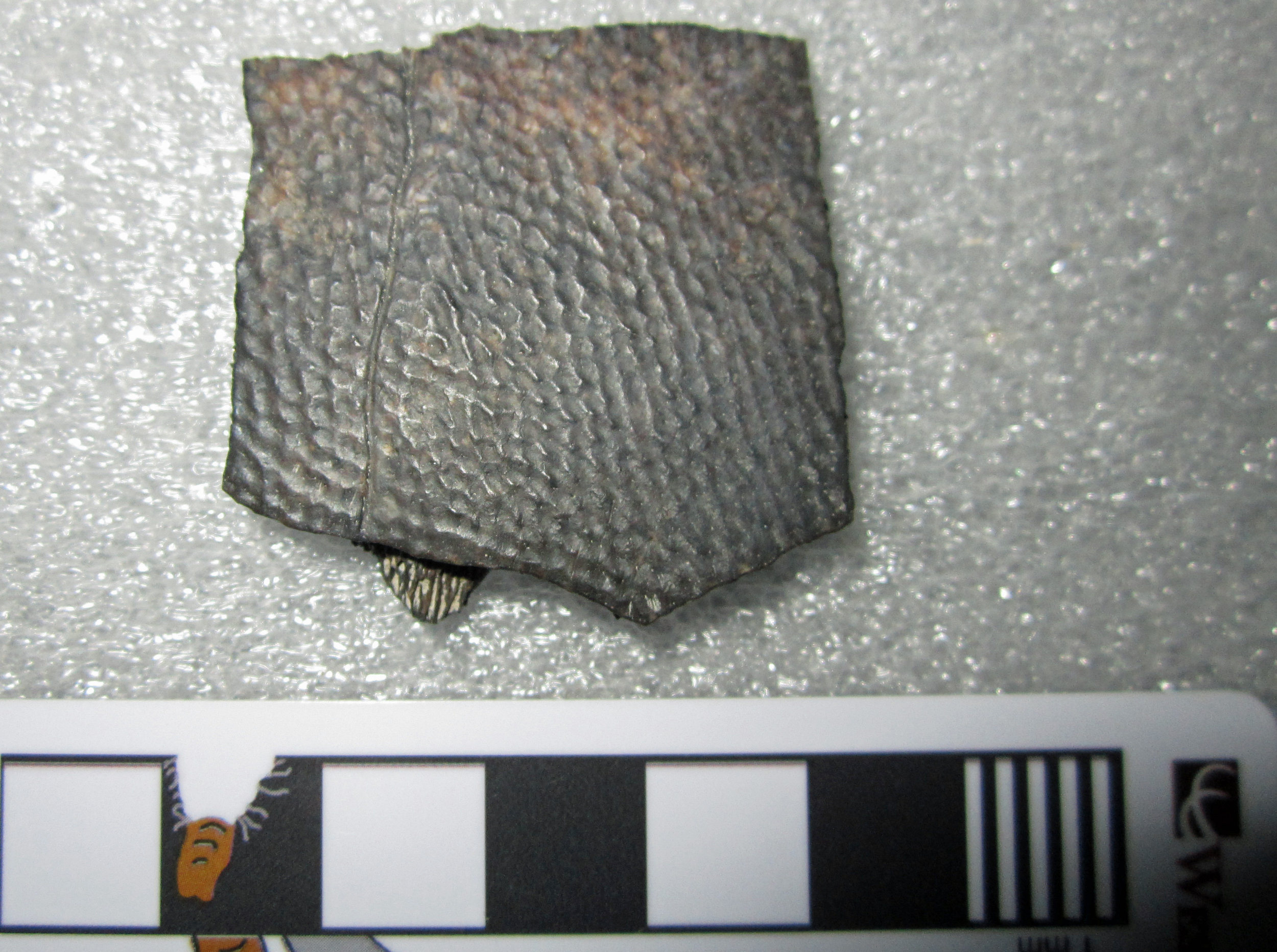
Fossil Friday - ceratopsian horn core
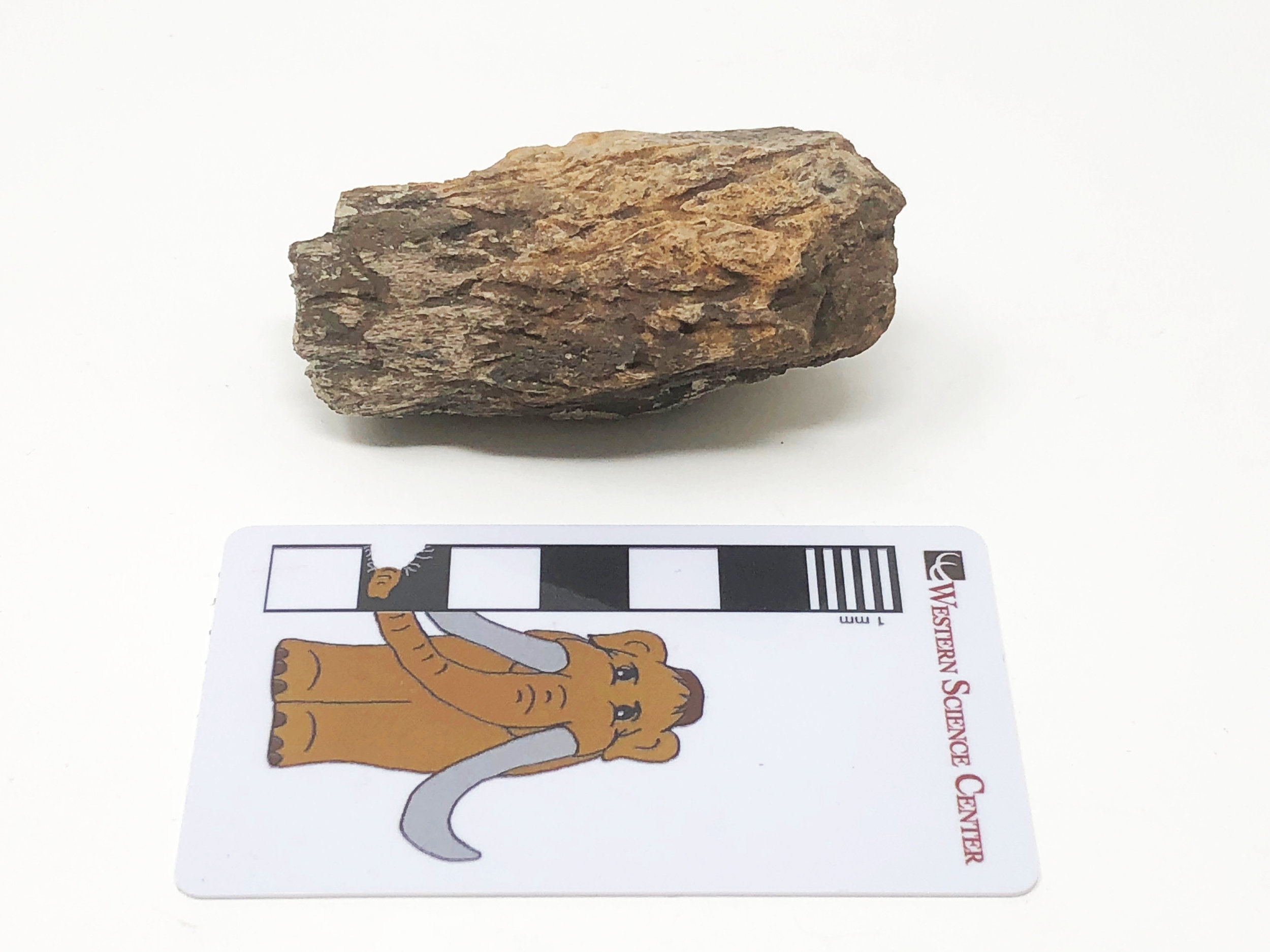 After a successful expedition earlier this summer, the Western Science Center crew returned to the Menefee Formation of New Mexico last week to search for more fossils of dinosaurs and other creatures that roamed the area 80 million years ago. As before, we were joined by colleagues from the Zuni Dinosaur Institute for Geosciences, students from the University of Arizona - Tucson, and volunteers from the Southwest Paleontological Society. Unlike the first trip, when we spent most of our time digging at two dinosaur quarries, this trip was mostly spent exploring for new fossil sites.This bone fragment is one of the pieces we collected while prospecting. The outer surface of this piece is riddled with deep branching grooves. This sort of texture is present only on the bony horn cores of ceratopsids, the big horned dinosaurs, such as Triceratops. When the dinosaur was alive, these grooves would have been occupied by blood vessels supplying the sheath of keratin (think fingernails) that covered the bony horn core. The photo shows the Menefee horn core fragment next to the brow horns of Ava, a horned dinosaur skull on display in WSC's "Great Wonders" exhibit. Ava's horns have the same texture.
 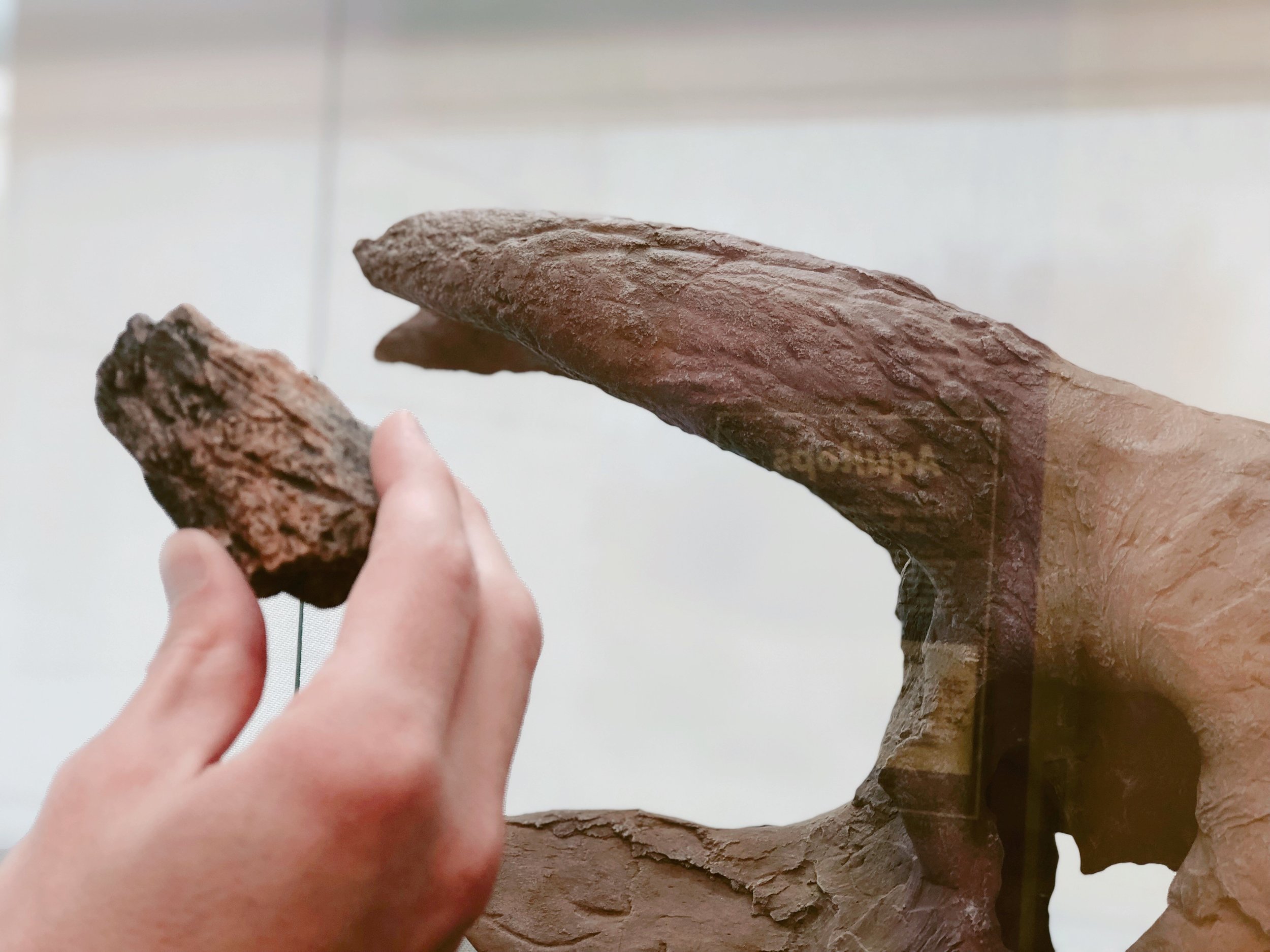 We will spend the next year preparing and studying the fossils we brought back from this summer's Menefee expeditions, and we will have much more to say about them in the very near future! Post by Curator Dr. Andrew McDonald
Fossil Friday - vole dentary
 While other projects have taken priority in recent weeks, we've still been making steady progress on our small but diverse collection of fossil mammals for Harveston, at the northern end of Temecula. I recently purchased a new camera, and tested its macro capabilities by making a photogrammetric model or a rodent jaw. These images are screenshots of the resulting 3D model after uploading to Sketchfab. At top is lateral view, and below is occlusal:
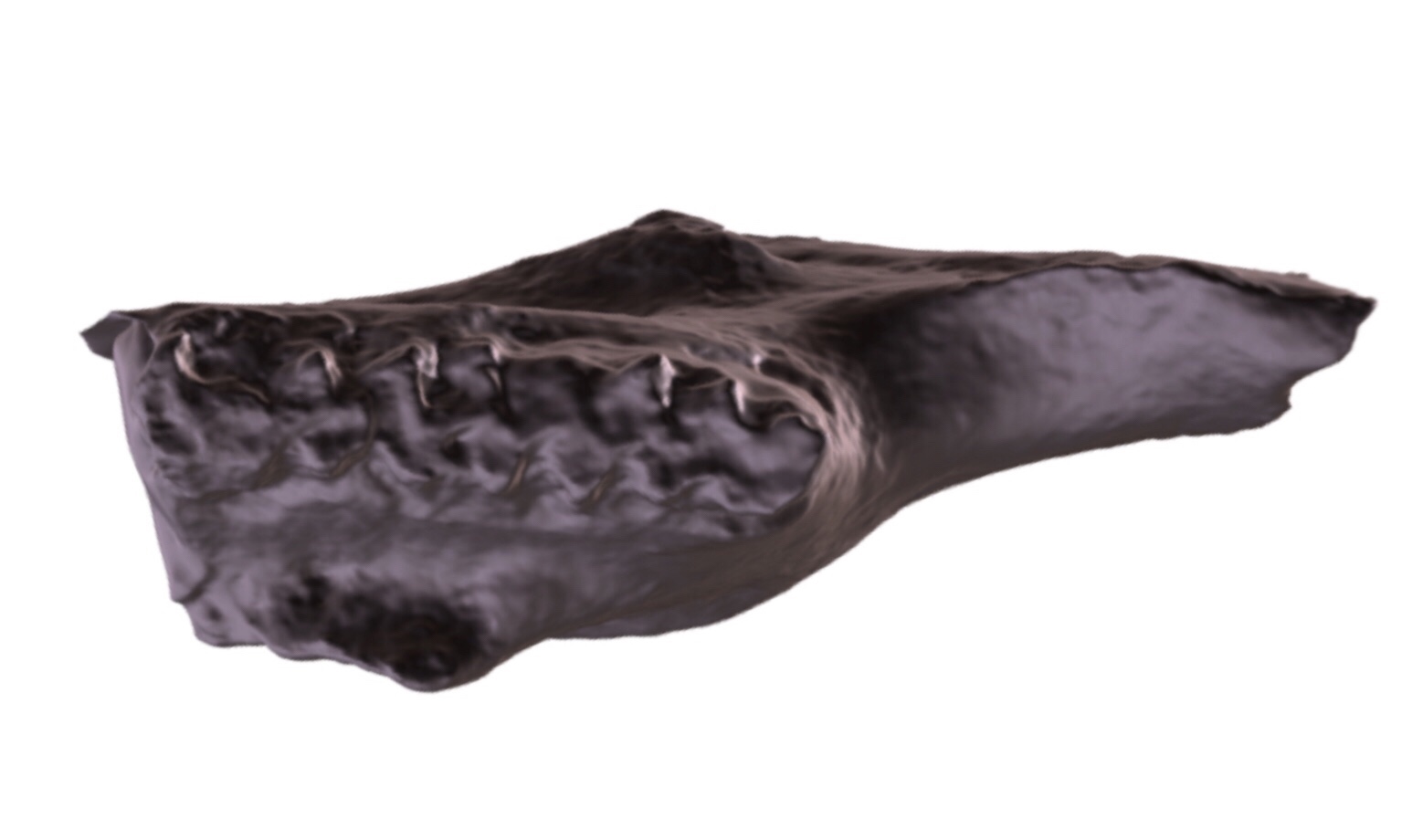 This is the anterior end of the right dentary. The 1st and 2nd molars are present, but the exposed part of the incisor is broken off, as is everything posterior to the 2nd molar. The enamel pattern on the occlusal surface of the teeth is typical of cricetid rodents, specifically voles of the genus Microtus. So far Microtus is the only genus of rodent we've positively identified from Harveston.The 3D model is available as a free download at Sketchfab: https://skfb.ly/6AHuK
Fossil Friday - a whale for WSC
 A few weeks ago, Western Science Center took delivery of the first fossil whale in our collection.This specimen was collected by PaleoSolutions during a construction project in Santa Cruz County. There are five separate field jackets, including one monster that's five meters long! That's the jacket shown above (notice the tiny scale bar on the right).This whale was found in Pliocene deposits, between 2 and 5 million years old. California has rich deposits of Pliocene whales, so when we've finished preparing this specimen (likely several years from now) there should be a lot with which to compare it. But even now, we can say a few things about it. First, this is a filter-feeding baleen whale from the suborder Mysticeti (hence the publicly-nominated nickname, Mystic). The only toothed whales (odontocetes) that get this large are sperm whales, and none of the visible cranial bones resembles the highly specialized elements in sperm whales. Below is a marked-up image with a few of the visible bones outlined (apologies for the lighting):
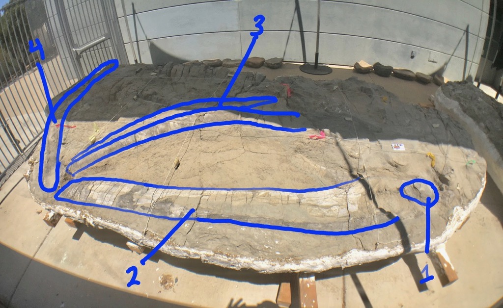 The bone labeled "1" is a vertebra, probably a thoracic or lumbar vertebra. This specimen includes numerous vertebrae, and all of them seem to have fused vertebral epiphyses, indicating that the whale is an adult.Number 2 is the left dentary; the left side of the lower jaw. It's not clear yet how much of the dentary is preserved, so it may be a bit longer than what is visible here. An interesting and important feature is the apparent absence of a coronoid process. The largest living group of baleen whales, the Balaenopteridae (including blue, fin, and humpback whales, among others) all have a large, prominent coronoid process. That means this specimen is more likely member of the right whale family (Balaenidae) or the grey whale family (Eschrichtiidae). (The weird pygmy right whale also lacks a coronoid process, and may not even be a right whale, but Mystic is not a pygmy right whale.)Number 3 I think are the premaxillae, which in whales extend from the tip of the upper jaw all the way to near the blowhole. These are strongly arched, which is consistent with both right whales and grey whales, but to me these look a bit more like a right whale.Number 4 I'm not sure about. I briefly thought is might be the broken continuation of the dentary, but I don't think that's right. The shape is wrong, and if you add that to bone 2 the dentary would be up in the blue whale-size range; Mystic isn't that big! I now think this might be the displaced and rotated maxilla. It should be running parallel to the premaxilla, so if it really is the maxilla it is way out of position, but this isn't unheard of. That shape is also consistent with a right whale maxilla.So, if this is a right whale (by no means a certainty), which right whale is it? There are actually only a handful of fossil right whale genera known. Of the four Pliocene genera, tiny Balaenella is only known from northern Europe, and is smaller than Mystic. Balaena includes the living bowhead whale as well as some extinct species. Eubalaena includes the three living right whale species, as well as several extinct ones. Most species of Balaena and Eubalaena are giants, quite a bit larger that Mystic (remember Mystic is an adult). I'm not ready to rule them out, but they seem unlikely. The fourth known Pliocene balaenid is Balaenula, a poorly understood genus that is known from all over the northern hemisphere, including California. Balaenula is a fairly small whale, and I think Mystic is likely a bit larger. But, if Mystic belongs to any right whale genus that has already been described, I think Balaenula is the best candidate.Mystic is on display in an outdoor preparation area at Western Science Center, so that the public can follow progress on its preparation in the years to come.
Fossil Friday - softshell turtle
 Turtles have a long and amazing fossil record, appearing around 260 million years ago, even before the earliest-known dinosaurs. By the latter most part of the age of dinosaurs, the Late Cretaceous Epoch, most of the modern turtle groups had evolved. Their fossils are extremely common in rocks that represent both freshwater and marine environments.Today's Fossil Friday is a piece of turtle shell that was collected by my colleagues and I in 2017 in the Menefee Formation of New Mexico. The shallow pits covering the surface suggest that it belongs to a trionychid - a softshell turtle, much like those that still live in North America today. This 80-million-year-old turtle was found at a diverse fossil site that also produced dinosaur bones, crocodile teeth, and large fish scales. These fossils were deposited in a freshwater environment, on a muddy floodplain near a slow-moving river.This is one of many turtle fossils discovered in the Menefee Formation by the Western Science Center, Zuni Dinosaur Institute for Geosciences, and Southwest Paleontological Society. We will have much more to say about them in the not too distant future! Post by Curator Dr. Andrew McDonald
Fossil Friday - hadrosaur tibia
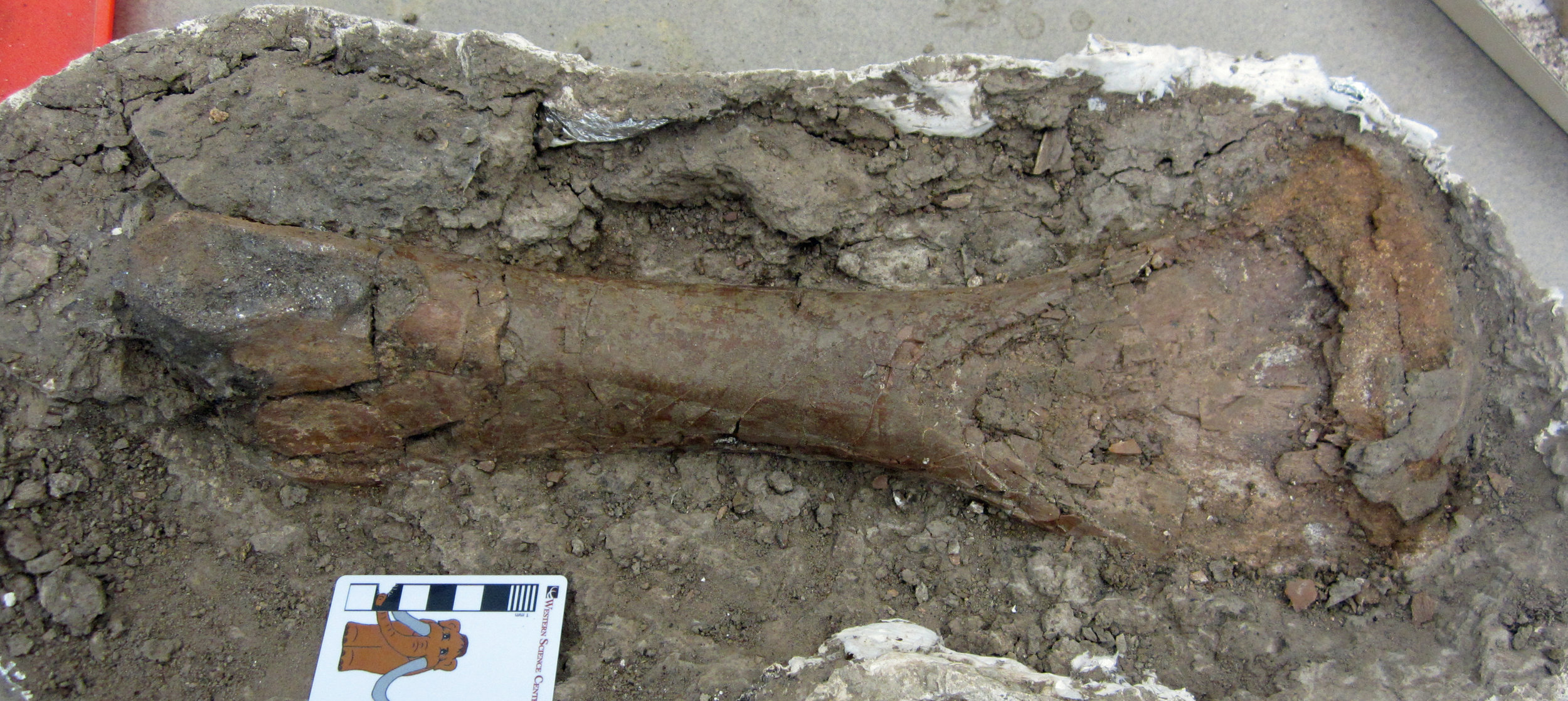 So far, most of the Late Cretaceous fossils I have shared with you for Fossil Friday have been from the Hell Creek Formation of Montana and were collected over a number of years by the late Harley Garbani. The Hell Creek dates to the very end of the age of dinosaurs, just before the mass extinction 66 million years ago.There are more Hell Creek fossils at WSC to share, but today I want to showcase another fossil from a somewhat older slice of the Late Cretaceous. Last month, I showed you some plant fossils from our field area in New Mexico. Today's fossil is the tibia of a plant-eating dinosaur, probably a young hadrosaur, one of the duck-billed dinosaurs. This bone was found by University of Arizona - Tucson paleontology undergrad Kara Kelley and is now being prepared at WSC by volunteer Joe Reavis.This bone was collected from the Menefee Formation of New Mexico, and is around 80 million years old. We are now preparing and studying this bone and many other fossils collected by the Western Science Center, our partners at the Zuni Dinosaur Institute of Geosciences in Springerville, AZ, and volunteers from the Southwest Paleontological Society. We had a very successful expedition to the Menefee Formation in May-June; you can read an account of it written by yours truly. And we'll undertake another round of field work in the Menefee in August!Post by Curator Dr. Andrew McDonald