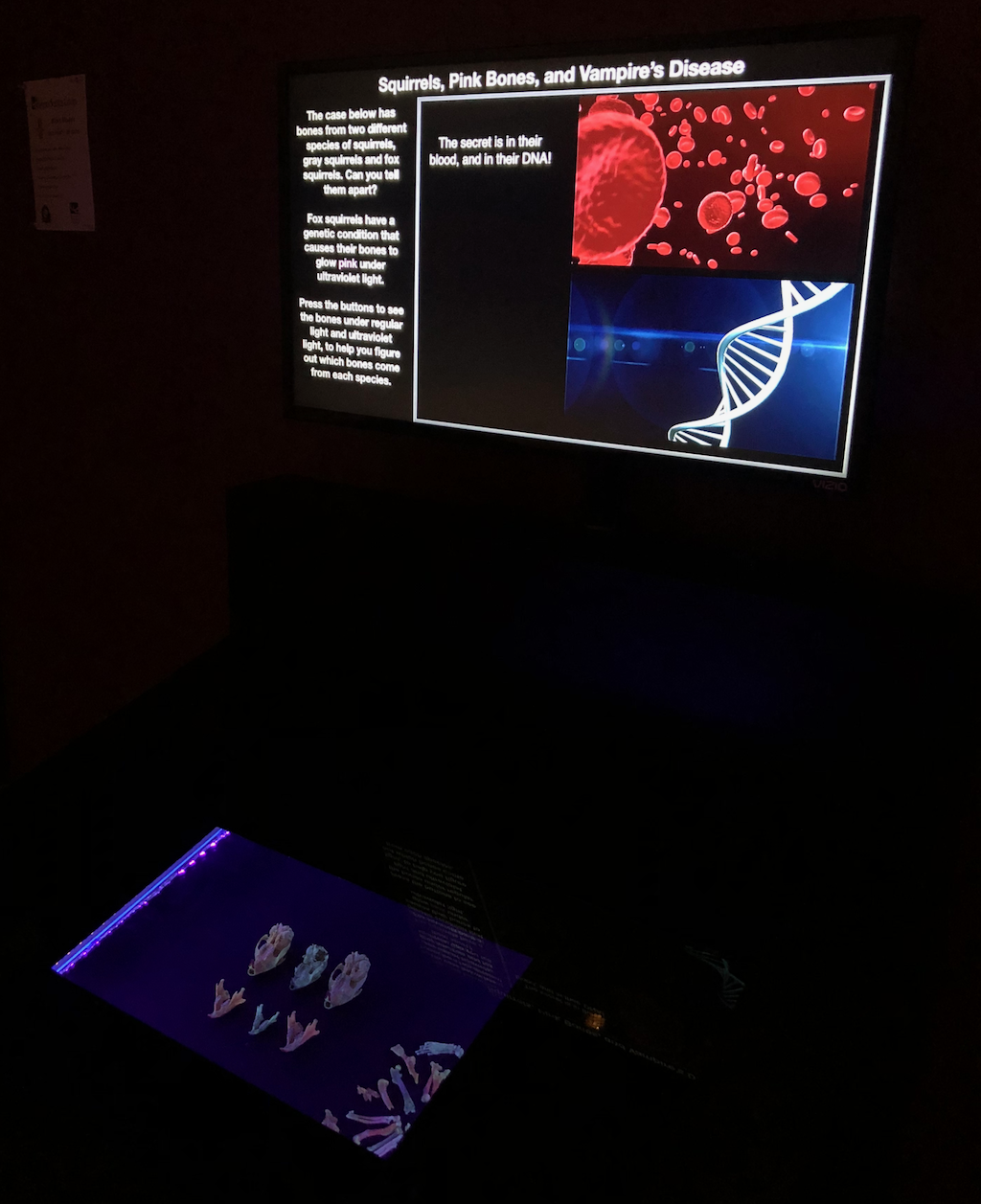
 This week we opened a new permanent exhibit at Western Science Center on eastern fox squirrels. This is based on research that I did in 2011-2013 in collaboration with Dr. Nancy Moncrief, Curator of Mammalogy at the Virginia Museum of Natural History (at that time I was Curator of Paleontology at VMNH). Much of the following is taken from a post I wrote for my old blog, Updates from the Paleontology Lab, on 29 February 2012.
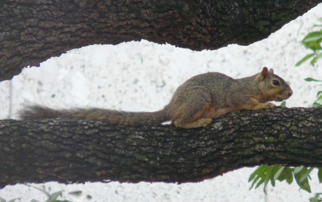 For many years, Nancy worked on the biogeographic patterns in the eastern fox squirrel, Sciurus niger (above), mostly by looking at marker genes. S. niger is an interesting species for this work because there are numerous subspecies that vary greatly in body size and coat color, as you can see in these specimens from the VMNH collections:
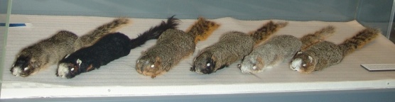 That’s a lot of variation! What makes it really interesting is that S. niger is a temperate species that doesn’t tolerate cold weather very well (they don’t hibernate). That means that their range was probably very restricted in the Pleistocene during the last glacial maximum, suggesting that all that phenotypic diversity may have arisen in less than 10,000 years.I was not involved in Nancy’s research, but because I do some biogeographic work Nancy would occasionally ask me to read her manuscripts or listen to her lectures to provide feedback from a paleontologist’s perspective. As a result I was somewhat familiar with her work.Then, in 2009 I was asked to write a book chapter reviewing the published record of vertebrate fossils in Virginia (which was finally published in 2016). While tabulating these species I noticed that, while the genus Sciurus had been reported from the Virginia Pleistocene, there were no reports of S. niger. Almost all the records were listed as Sciurus sp.Realizing that being able to tell when fox squirrels show up in different places could be useful for Nancy’s work, I asked her about the lack of definitive species-level identifications. The problem, she explained, is that it’s really difficult to distinguish S. niger from the closely related, sympatric, eastern gray squirrel (Sciurus carolinensis, below) when only the skeleton is available.
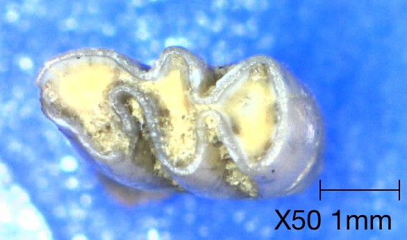
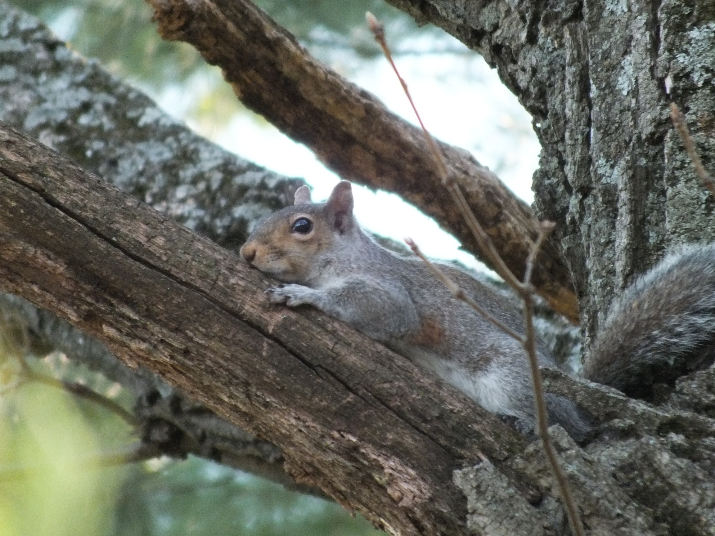 The problem is that tree squirrels are pretty conservative in their skeletal morphology, especially in the postcranial skeleton, with few changes since the Miocene at least. The average size and maximum size of fox squirrels are greater than in gray squirrels. However, there is considerable overlap; the largest gray squirrels are bigger than the smallest fox squirrels. So using body size for identification only works for really big specimens.There are also some detail differences in the skull and mandible, but they tend to be somewhat inconsistent. Easily the most reliable feature is the presence of a rudimentary upper 3rd premolar in S. carolinensis, which is absent in S. niger. Below is the upper right tooth row of S. carolinensis, top, and S. niger, bottom. In each specimen anterior is to the right. Note the tiny, peg-like premolar at the front of the tooth row in S. carolinensis only:
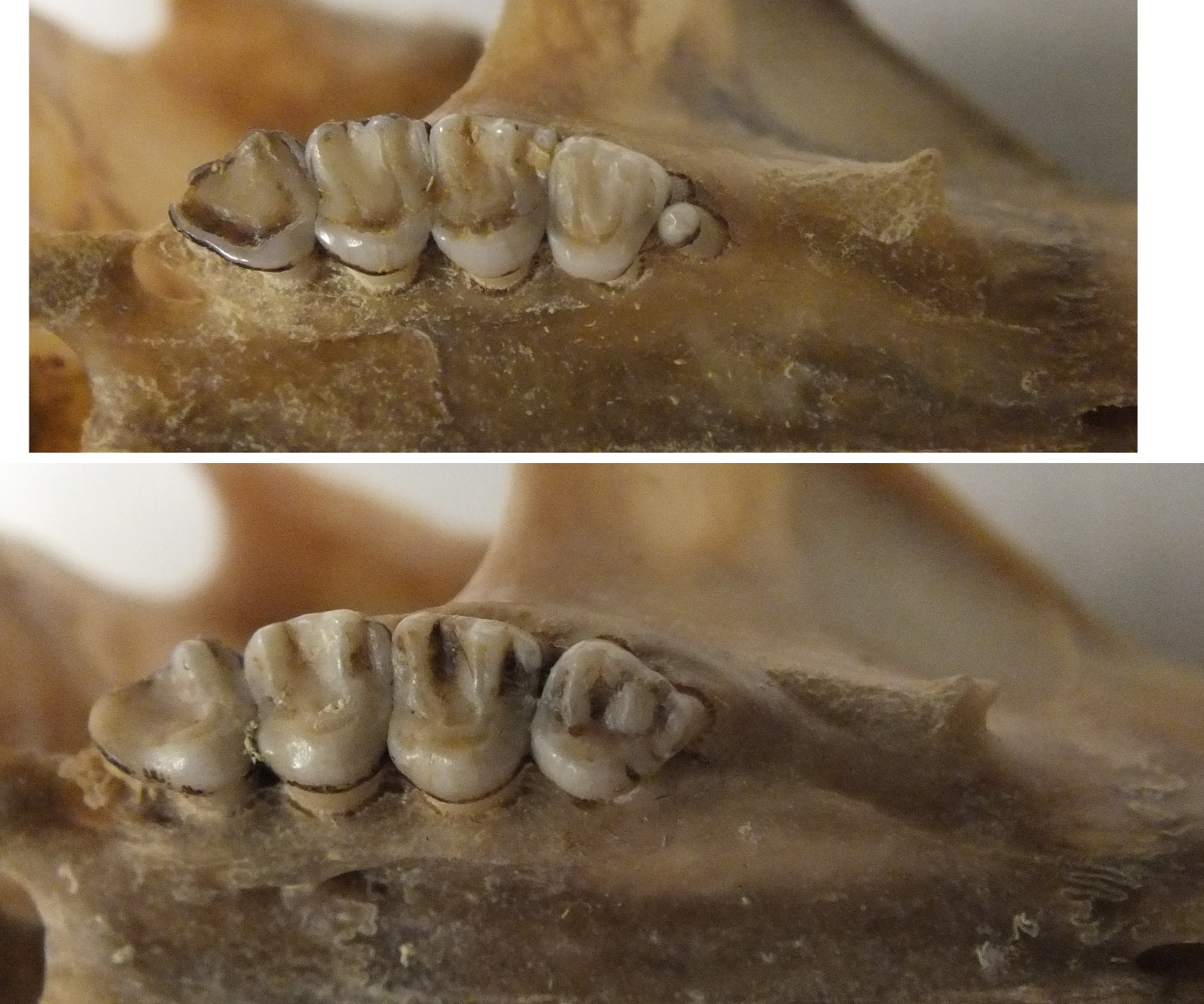 Unfortunately, it’s pretty rare to preserve that particular part of the maxilla, so the extra premolar is of limited use in identifying isolated bone fragments in Pleistocene deposits. In an irrational fit of optimism, I suggested to Nancy that I might be able to come up with features for distinguishing between the two species based on isolated material, especially since the VMNH collections include hundreds of modern squirrel skeletons to use as references.After several frustrating days looking at squirrel bones, I was ready to give up. There simply don’t seem to be any consistent features for distinguishing postcranial remains other than size, which is unreliable. Then, as Nancy and I sat together discussing squirrel trivia, she asked if I knew that fox squirrel bones glow pink under ultraviolet light. It hit us both at the same time; only fox squirrel bones fluoresce under UV light! Here’s an S. niger skull under visible light (left), and the same skull under UV light (right):
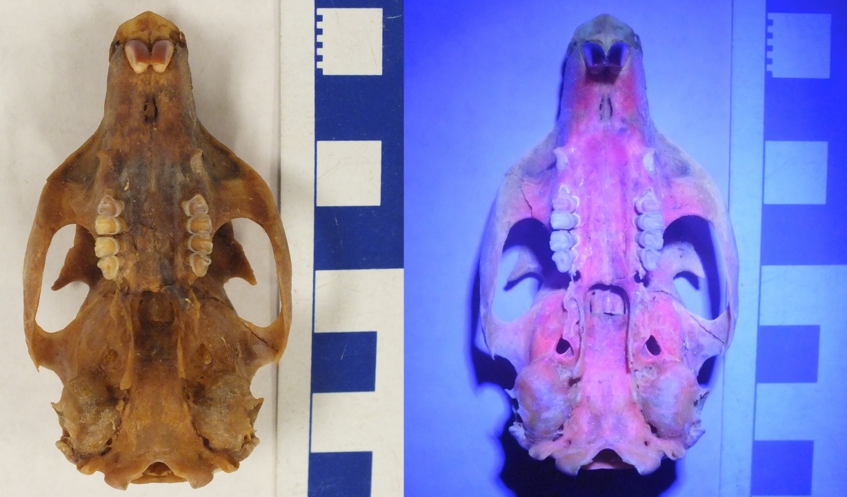 So here’s the deal: eastern fox squirrels almost universally have a genetic condition called congenital erythropoietic porphyria (CEP). This is caused by a mutation in one of the genes involved in the heme chain (heme is a key component of hemoglobin). Specifically, animals with CEP can’t properly metabolize the enzyme uroporphyrin, which as a result starts to build up in the urine, bones, and teeth. Uroporphyrin fluoresces pink when exposed to ultraviolet light.As I mentioned above, CEP is normally not present in gray squirrels, so we can potentially use the presence of fluorescence to distinguish between fox and gray squirrels, even when there’s a size overlap. For example, the skulls below are organized by increasing size from left to right. Which ones are S. niger, and which ones are S. carolinensis?
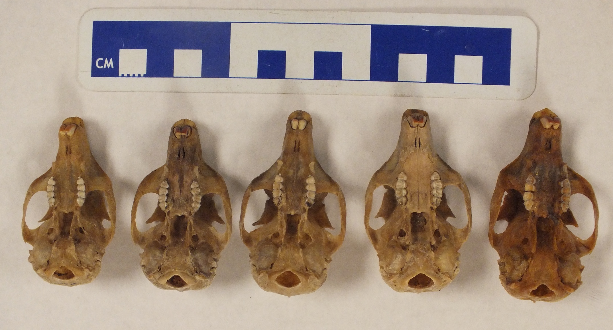 Here are the same skulls under UV light:
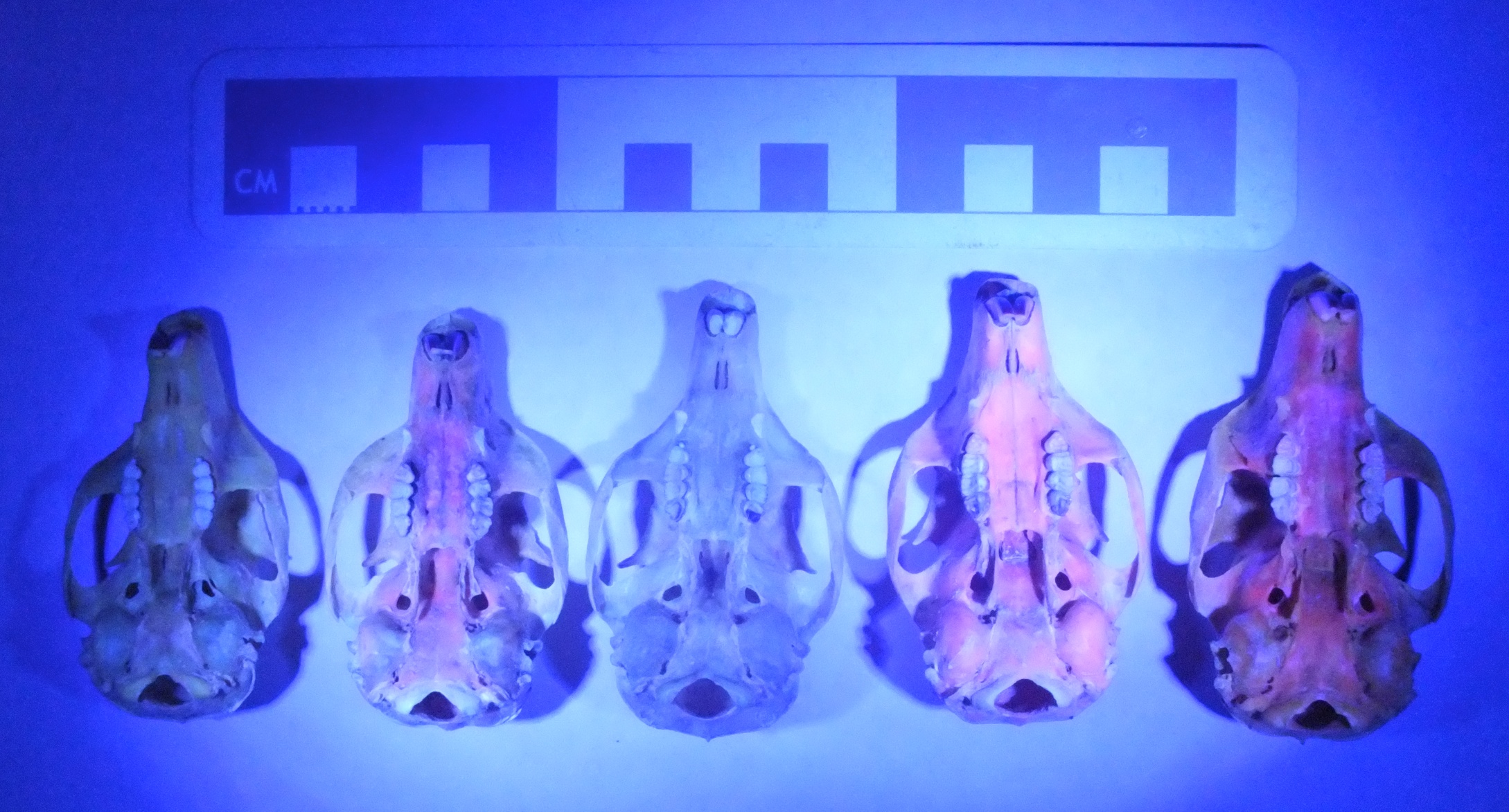 The 2nd, 4th, and 5th skulls all fluoresce, and are all fox squirrels.CEP has been reported as a rare condition in several different mammal species, including humans. It’s typically a pretty dreadful condition that results in photosensitivity, skin lesions, and anemia among other problems. The combination of sensitivity to sunlight and anemia (requiring blood transfusions) has led to CEP sometimes being called "Vampire's disease". For some unknown reason, eastern fox squirrels don’t seem to suffer from the adverse effects of CEP even though they have elevated uroporphyrin levels.Biologists have known since the 1930’s that fox squirrels have fluorescent bones (Turner, 1937), and that sometimes they’re even pink under visible light. There were a few studies in the 1970’s looking at the condition in some detail (Levin and Flyger 1971, for example), but somehow this knowledge was never applied to fossil remains. The question, of course, is that since a particular protein is responsible for the fluorescence, would it still be detectable in ancient specimens?The Florida Museum of Natural History lists several specimens of S. niger among their Pleistocene/early Holocene collections, and they happened to be the host for the 2011 SeAVP meeting. The day before the meeting I spent a few hours in their collection with a tray of squirrel bones from Devil’s Den Sinkhole and a UV light. Sure enough, even though these bones have been sitting at the bottom of a sinkhole (that’s currently flooded) for at least 7,000 years*, several of the bones still fluoresce. The best example was this partial skull:
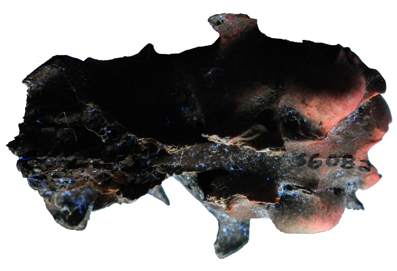 There’s definitely a high “coolness” factor here; we diagnosed a genetic condition in a late Pleistocene/early Holocene squirrel! But in addition to that, we’ve given ourselves a pretty powerful tool for examining ancient squirrels. To be sure, there are limitations: the fluorescence seems to disappear if the bones have been mineralized, and the condition may not be truly universal in S. niger. Therefore, this only works as a positive test for the presence of S. niger; the absence of fluorescence doesn’t definitively tell us anything. But on the other hand, this allows us to identify even fragmentary postcranial remains, the test is non-destructive and inexpensive, and the results are immediate. And it’s spectacularly easy to use on mixed bone deposits, including samples obtained by screening. Here’s a sample of bones from multiple taxa obtained by screening a sediment sample from a late Holocene site:
 And here’s the same sample under UV:
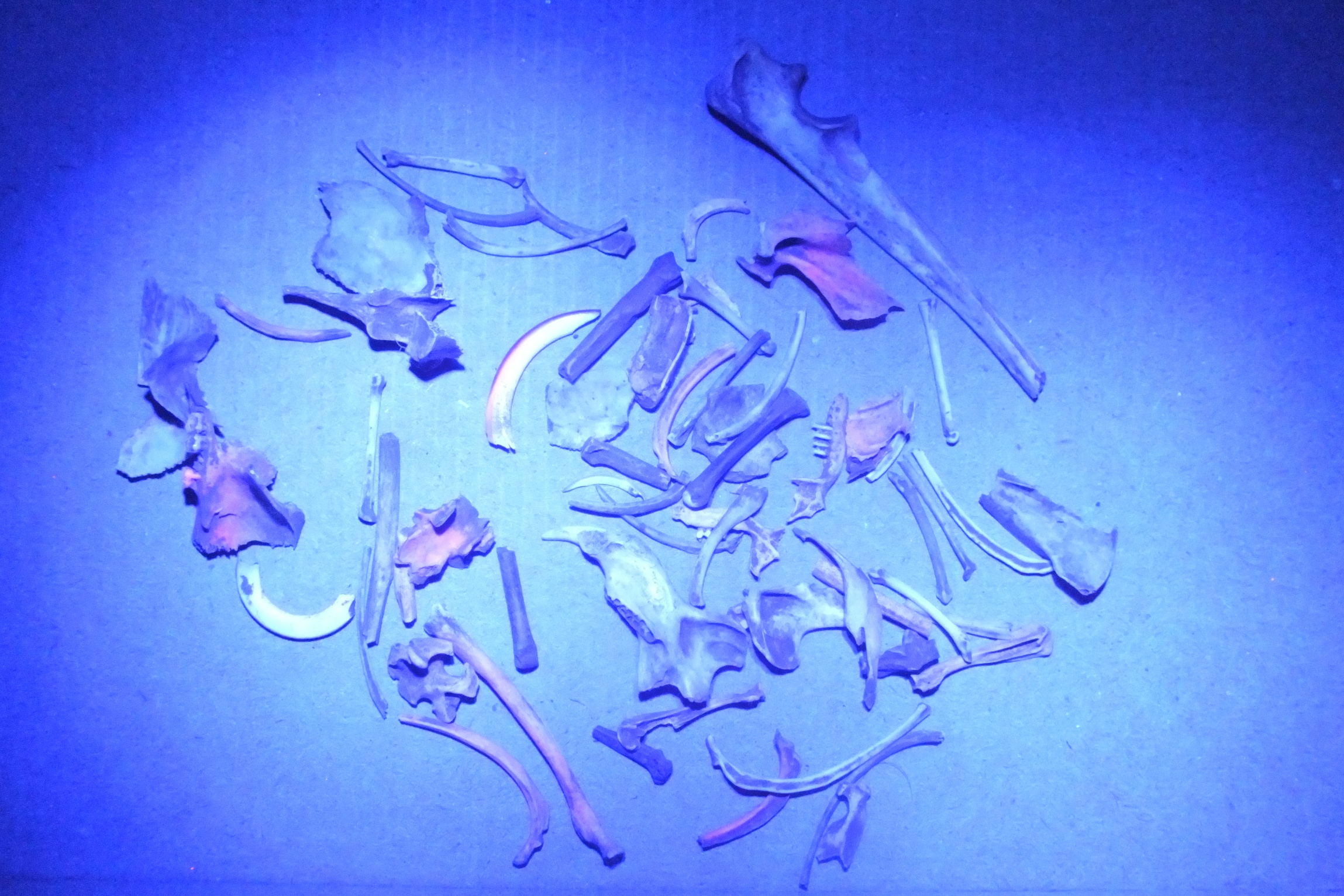 A dozen of these bones turned out to be fox squirrel (some of them are hard to spot in a photograph). They include difficult-to-identify elements like ribs; notice the two at the bottom center of the photo.Nancy and I published the results of our study of the Devil's Den fossil squirrels in 2012. In a follow-up paper in 2013, we examined squirrel remains from Native American archaeological sites in Virginia that were roughly 1,000 years old. Even though these specimens had in most cases been burned, they still fluoresced, as with this tibia:
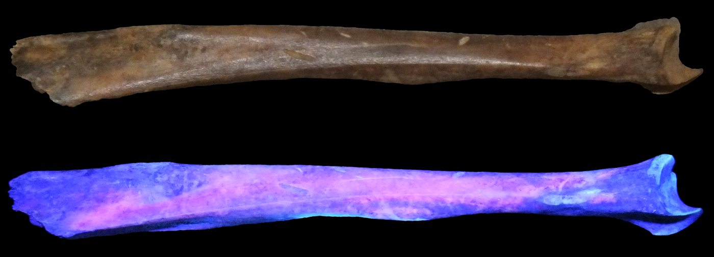 This research brought up a lot of additional questions that we never fully addressed. We found, both in the literature and from our own observations, that not all fox squirrels fluoresce equally, and a minority don’t seem to fluoresce at all. Why? Are some populations more prone to CEP, is there an ontogenetic component to uroporphyrin levels, or is there just a significant amount of individual variation? Moreover, certain elements seem to fluoresce particularly well (for example, the maxillae, the dentaries, the incisors, and the 4th upper premolars) while others often don’t fluoresce at all (like the nasals and the molars). Since moving to California, I've also learned that there is an introduced population of Eastern fox squirrels in Los Angeles County, which have been isolated from other S. niger populations now for toughly 100 years. I've not yet had the chance to examine one of these squirrels, but I wonder if their bones still fluoresce?*There’s some uncertainty about the age of the Devil’s Den deposits. They were published as being approximately 7,000 years old, but the presence of extinct animals such as mastodonts in the same deposit indicates that it is more likely late Pleistocene (Martin and Webb, 1974).References:
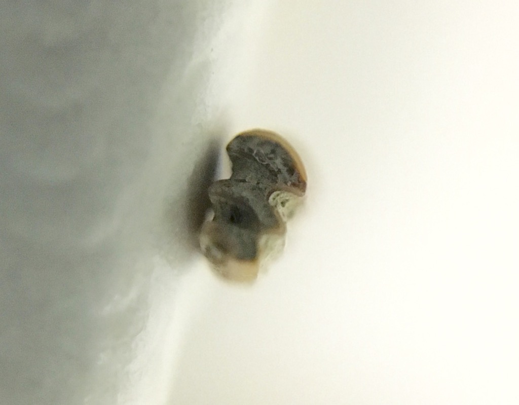
 Abstract deadlines are coming up for conferences taking place in the first 6 months of 2019, so we're trying to collect as much data as possible for our submissions. One of these is a follow-up to our abstracts from last year on the Harveston locality in Temecula, but this time looking at the small animals ("microvertebrates"). One of our high school volunteers (or "Max's Minions", as we call them), Charlotte Hohman, is taking the lead on this project, sorting vials of tiny rodent, lizard, and bird bones and trying to identify them. This week we may have found a bone from a relatively uncommon mouse, the most metal mouse in Southern California.The bone in question is a left calcaneus (heel bone). The image isn't the greatest because it was taken by holding an iPhone up to a microscope eyepiece; we just wanted a quick reference image. The total length is about 5 mm.After comparing this to references, we're pretty sure this calcaneus comes from a southern grasshopper mouse, Onychomys torridus. If correct, this is our first grasshopper mouse from Harveston. Onychomys is known from the Diamond Valley Lake fauna, but is quite rare compared to the other rodents (in fact, I think we have more mastodons than grasshopper mice!).So this is just a mouse; what makes it metal? Unlike their close relatives such as deer mice, which are omnivores leaning toward herbivory, grasshopper mice are voracious predators. They primarily eat insects and other arthropods, but will eat other mice on occasion.They are known especially for eating bark scorpions. Apparently, when stung by a scorpion, the mouse's pain receptors shut down; even though they may get stung multiple times, they don't feel the pain. Northern grasshopper mice will attack centipedes, which are also venomous; the mouse is just fast and sting (I haven't been able to confirm if southern grasshopper mice also eat centipedes).And, if all that isn't enough, grasshopper mice are territorial. But their territories can be over 20 acres (!), and they'll warn away interlopers by
 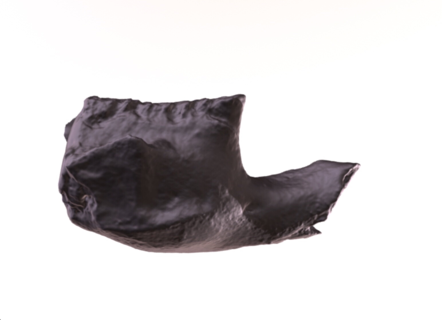 While other projects have taken priority in recent weeks, we've still been making steady progress on our small but diverse collection of fossil mammals for Harveston, at the northern end of Temecula. I recently purchased a new camera, and tested its macro capabilities by making a photogrammetric model or a rodent jaw. These images are screenshots of the resulting 3D model after uploading to Sketchfab. At top is lateral view, and below is occlusal:
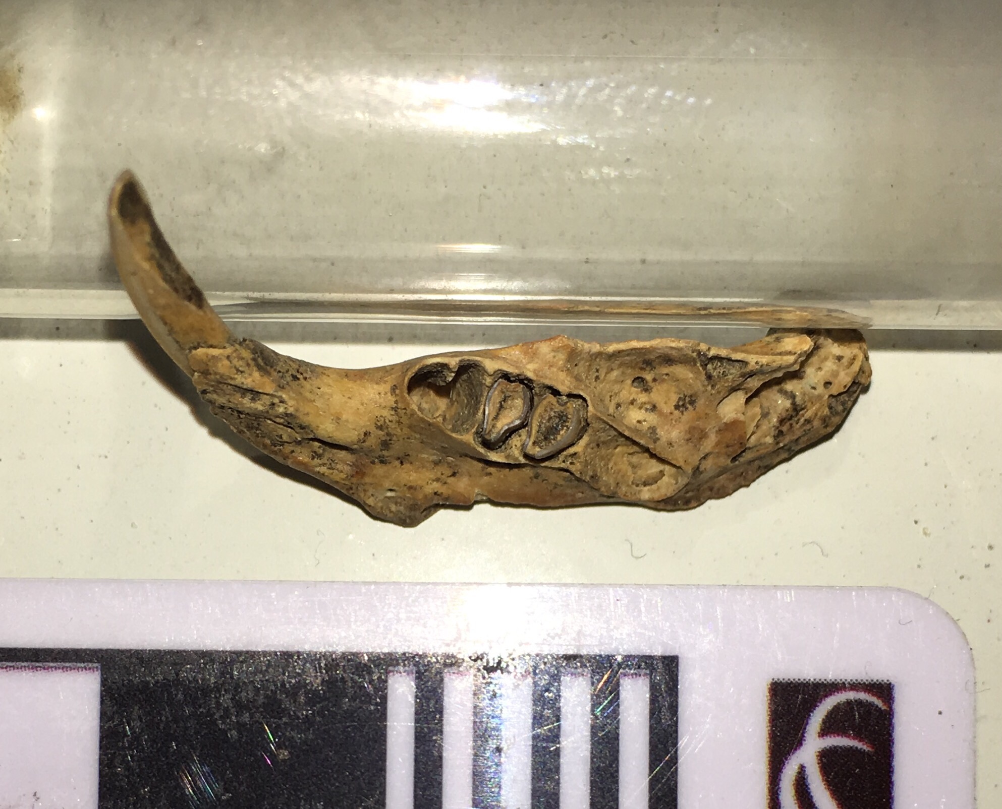
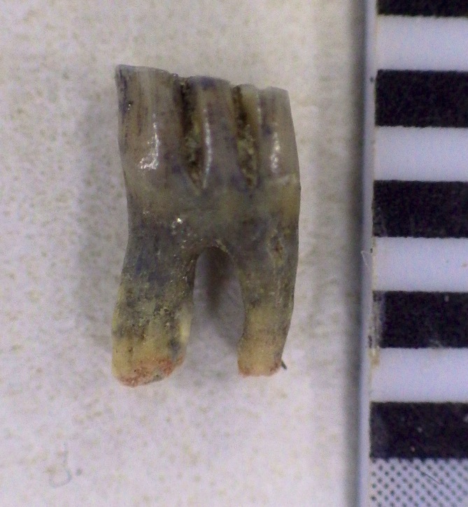
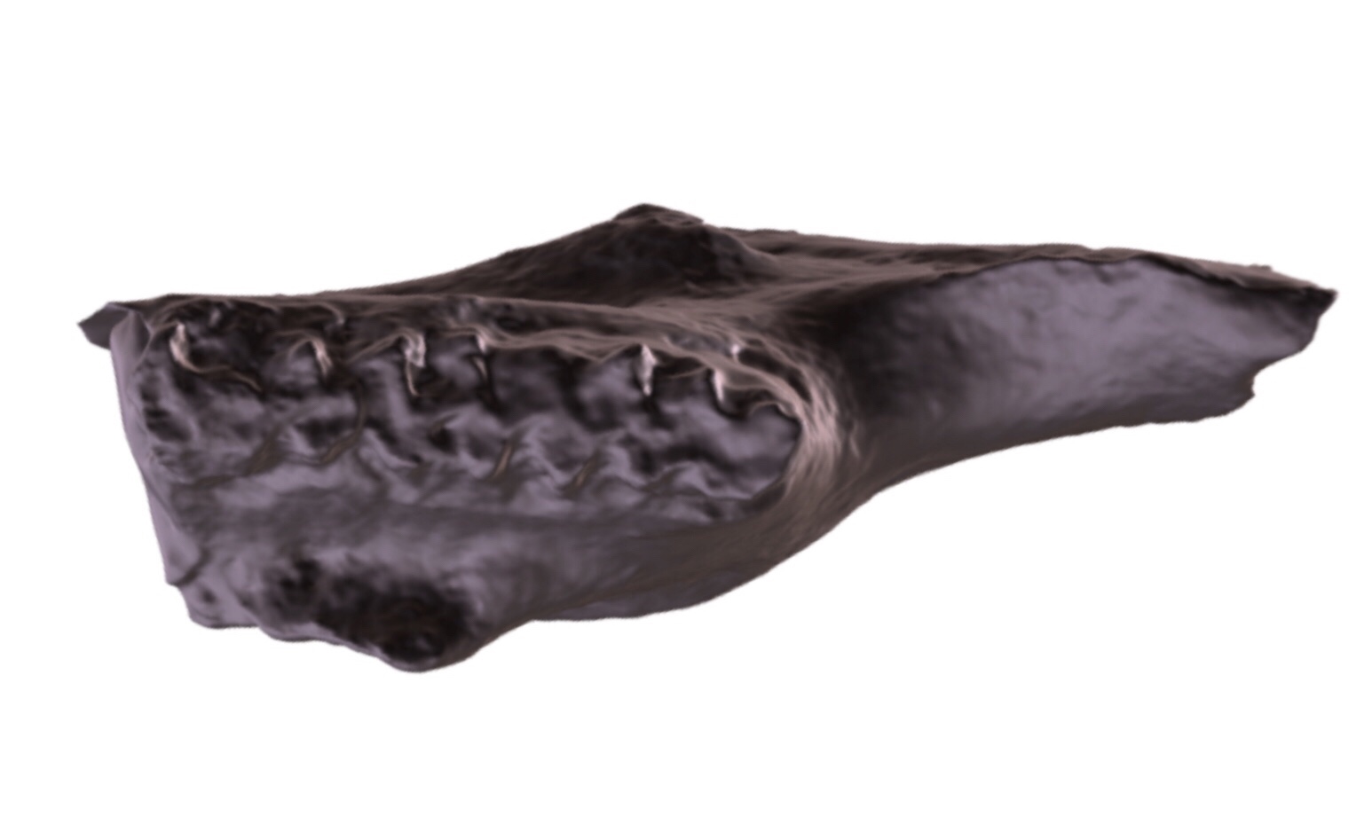 This is the anterior end of the right dentary. The 1st and 2nd molars are present, but the exposed part of the incisor is broken off, as is everything posterior to the 2nd molar. The enamel pattern on the occlusal surface of the teeth is typical of cricetid rodents, specifically voles of the genus Microtus. So far Microtus is the only genus of rodent we've positively identified from Harveston.The 3D model is available as a free download at Sketchfab:
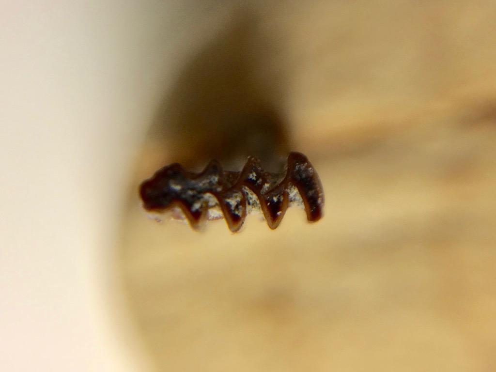
 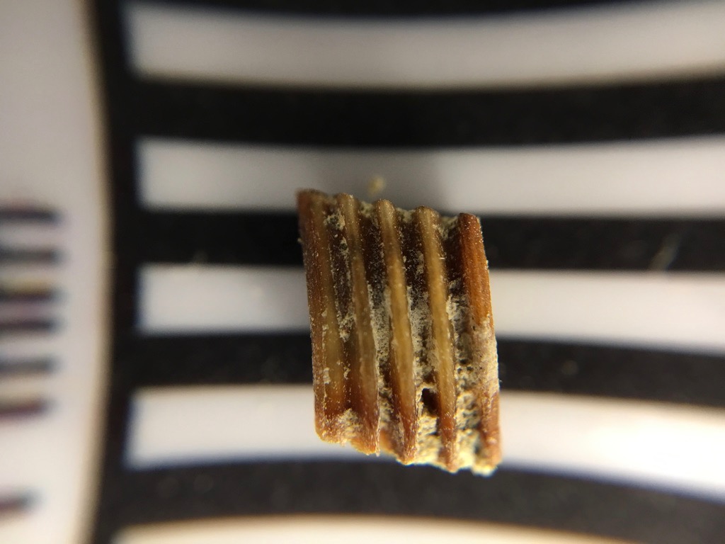 This week is National Rodent Awareness Week. Fossil rodents may not get a lot of headlines, but they are often the most common vertebrate fossils in Neogene terrestrial deposits, and have the potential to convey huge amounts of information about age and paleoenvironment.One of the larger collections at the Western Science Center is from the Southern California Edison El Casco substation in Riverside County. These Late Pliocene/Early Pleistocene deposits of the San Timoteo Formation produced over 16,000 fossils, including large numbers of rodents.Shown above is the right lower first molar of Microtus, a vole. It's shown in medial view, with anterior to the left. The black and white bands under the tooth are each 1 mm thick, so the whole tooth is about 4 mm tall.Voles and other members of the subfamily Arvicolinae (including hamsters, lemmings, and muskrats) are herbivores, and often eat abrasive food such as grasses. Like many other herbivores, this is reflected in their tooth anatomy, which includes loops of enamel with exposed dentine in the middle. Because the enamel is harder than the dentine, it always sticks up a bit higher than the dentine and allows the tooth to stay sharp even as it wears down. The enamel loops are expressed as ridges or columns when seen from the side, and are clearly visible in the view above. In occlusal view (below), the loops are expressed as a series of alternating triangles, a pattern that is characteristic of the arvicolines.