

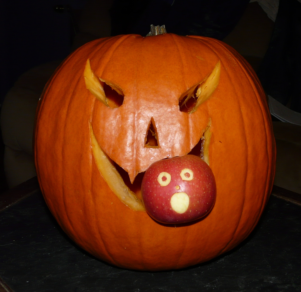
 Pumpkins are an interesting fruit. Curcurbita pepo is one of several domesticated species of the genus Curcurbita, vines that are native to the Americas. Curcurbita is a ecologically diverse genus, with some species needing a continuous water supply while others can live in arid conditions, so it is found natively in a variety of habitats. The fruits, which are technically berries, generally have a thick rind with a softer interior where the seeds are located. In most species the rinds are bitter, but the interior is often more palatable and rich in nutrients. As a result it became one of the first domesticated plants in North America more than 8,000 years ago.With large, nutritious fruits, we can be confident that wild Curcurbita were being eaten by more than just humans. In 2006 Lee Newsom and Matthew Mihlbachler published a detailed report about an American mastodon dung deposit in northern Florida. The vast majority of the dung consisted of small cypress twigs, chopped up and stripped of bark, but there were other plant remains from at least 57 species. Among their samples were 156 Curcurbita seeds, showing that this was a popular item on the mastodon menu. Using the Newsom and Mihlbachler paper as a guide, a few years ago we made a simulated piece of mastodon dung for exhibit at WSC, including numerous pumpkin and squash seeds:
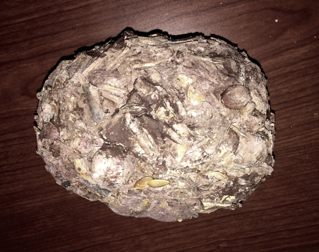 Newsom and Mihlbachler also noted that, while Curcurbita seeds were relatively common in their sample, rind fragments were almost completely absent. It seems that mastodons may have disliked the bitter rinds just like we do, and broken the gourds open to get at the interior. Captive elephants that are fed pumpkins seem to have discovered the same trick:[youtube https://www.youtube.com/watch?v=2CzXH2Eg1hw&w=560&h=315]So far no Pacific mastodon coprolites have been identified, but hopefully one day we'll be able so say as much about their dietary habits.Reference:Lee Newsom and Matthew C. Mihlbachler, 2006. Mastodon (Mammut americanum) diet and foraging patterns based on analysis of dung deposits. Chapter 10 in S. David Webb (ed.), First Floridians and Last Mastodons: The Page-Ladson Site in the Aucilla River, Springer, p. 263-331.
Fossil Friday - traces on a mammoth rib
 Organisms don't exist in a vacuum. The whole concept of an ecosystem emphasizes the interactions between an organism and its environment, including with other organisms. A large organism like a mammoth can have wide-ranging effects on numerous other organisms, even after its death.The specimen shown above is a partial Colombian mammoth rib from Diamond Valley Lake, currently being prepared by volunteer Tim Dooley. We have a large number of rib fragments such as this one, and at first glance they all look pretty much the same. But under close examination they often tell unique stories.After removing glue used to stabilize the specimen in the field, we realized this bone has a number of small, circular pits covering its surface:
 There are also some rough, pitted areas:
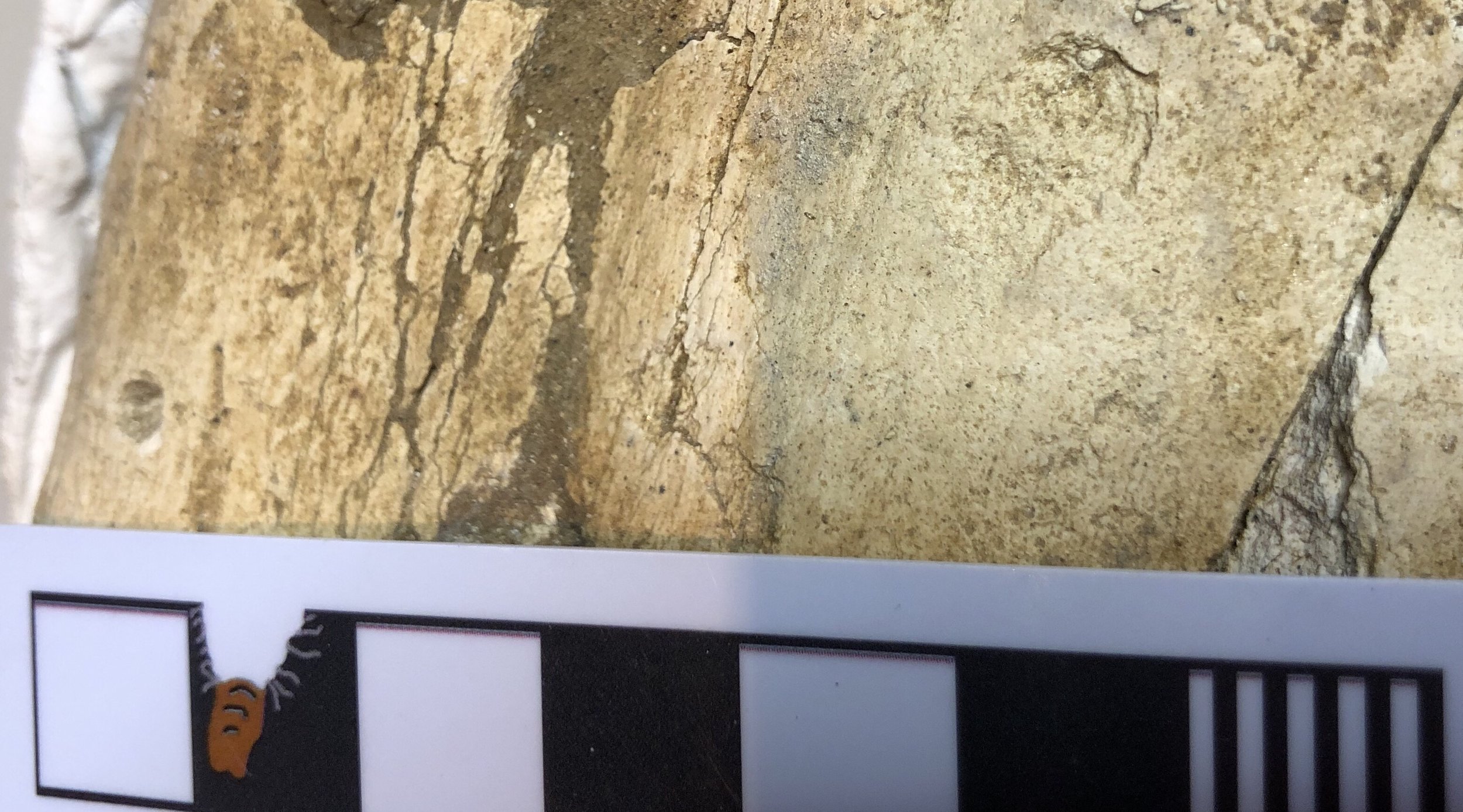 While I'm not 100% sure, I believe these are traces made by scavenging insects or other invertebrates, especially the rough patches in the last photo. I'm a little less certain of the circular holes. They are reminiscent of vertebrate bite marks, but they aren't paired and are tiny (all are less than 5 mm in diameter), so I think some sort of burrowing or bone-eating invertebrate is more likely.Marks such as these are quite common on DVL bones, and may eventually give us some insight into the invertebrate fauna from the valley, which left us almost no body fossils.
Fossil Friday - camel elbow
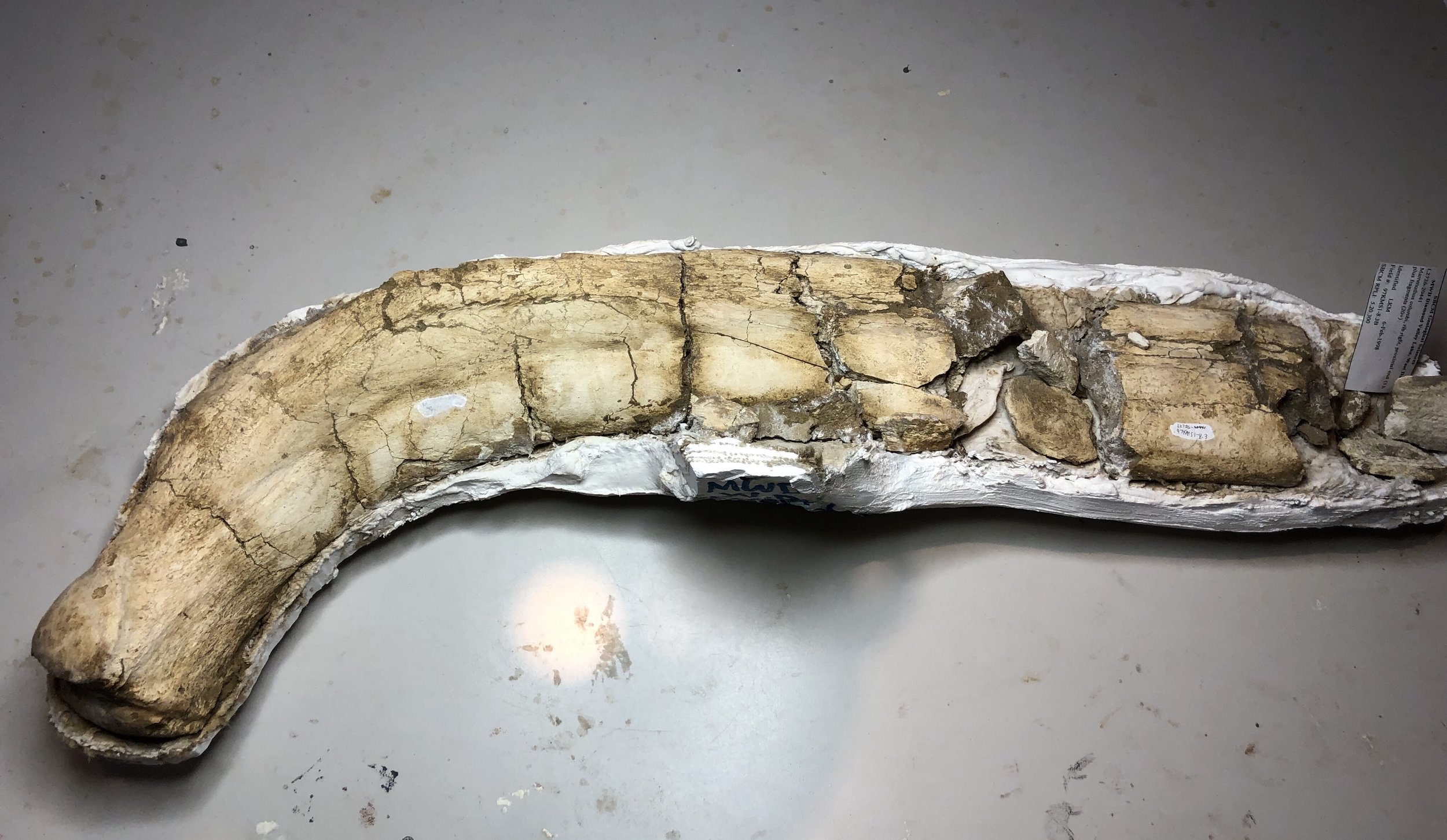
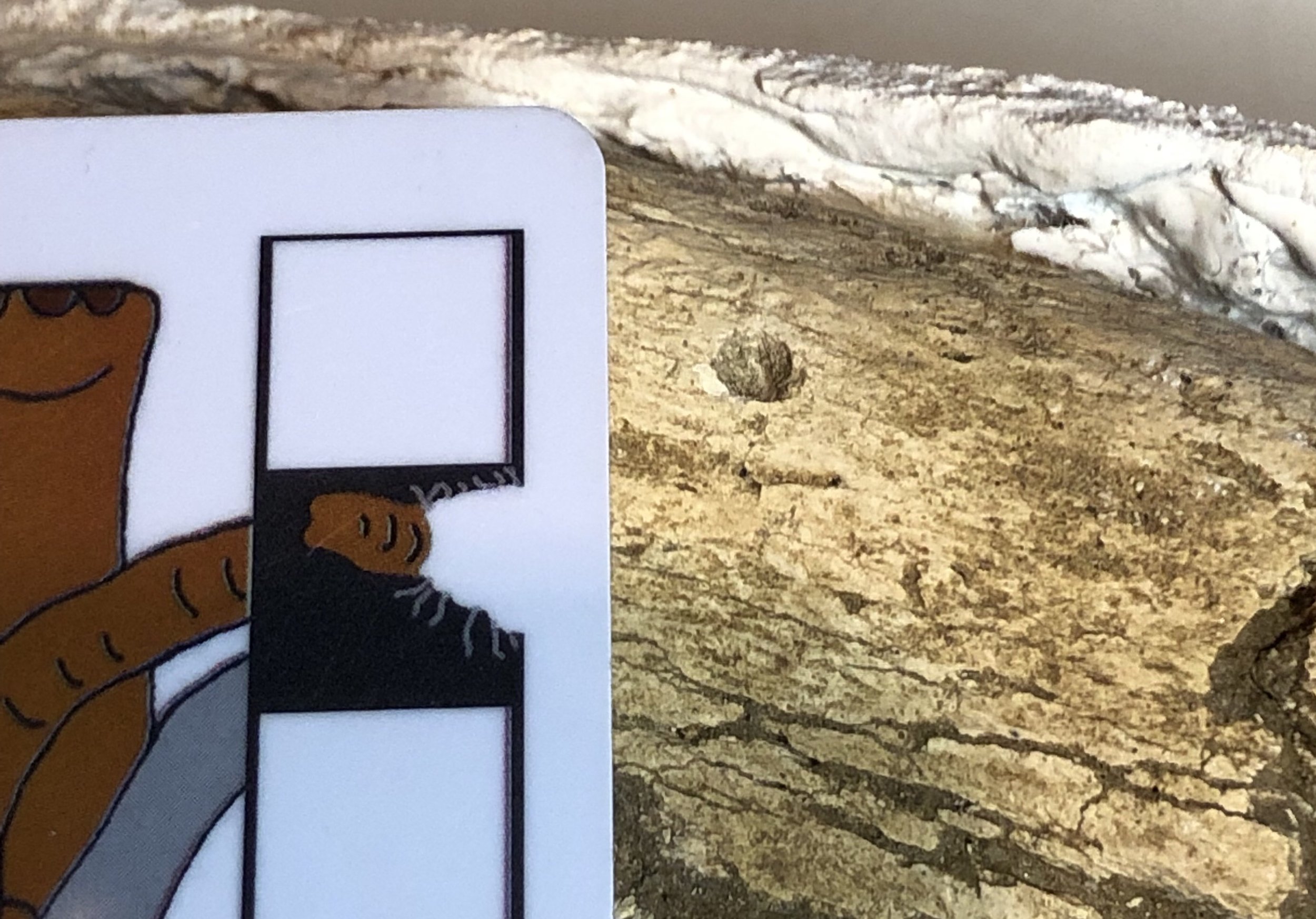
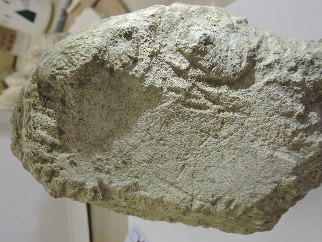
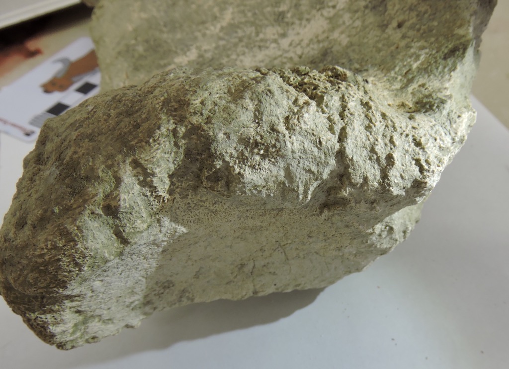
 We're continuing our focus on Pleistocene fossils from Murrieta, California this week with a single bone fragment that has a lot going on.This bone is labeled in our collections as an ulna from Equus, a horse. It is indeed part of a left ulna, one of the bones from the forearm. More specifically, it's the olecranon process, the proximal end of the ulna that forms the point of the elbow. The triceps muscle attaches to the olecranon process, allowing you to straighten your arm (or front leg, in this case). However, after spending several hours comparing this to various publications and our Diamond Valley Lake collections, I'm not convinced it's a horse.For an olecranon process of this size, there are really only three likely animals from the Pleistocene of Southern California it could belong to: horse, camel, and bison. Everything else is either much larger (mammoth, mastodon) or much smaller (mule deer). (OK, to be fair, short-faced bears and ground sloths are in this size range, but their ulnae look nothing like this.) Horses, camels, and bison are all known from this site, but while it's a little on the small side, this bone is the best match with the western camel, Camelops hesternus.There is another interesting feature on this bone. Notice the four circular depressions near the tip (on the right). Here's a closeup:
 There are a few comparable depressions on the other side:
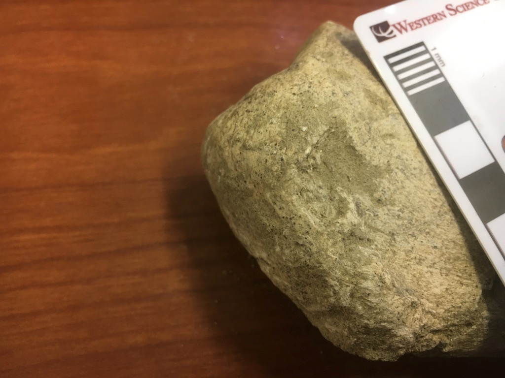 These appear to be bite marks, from some carnivoran chewing on the end of the bone. There are several other scrapes that appear to be gnaw marks. So far we have not identified any carnivoran bones from this site, but they certainly made their presence felt.We have scanned and 3D-printed this bone (print shown below with the original), and the scans can be viewed on Sketchfab at https://skfb.ly/6xNMM.
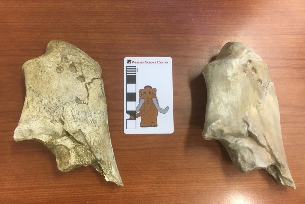
Fossil Friday - proboscidean ulna
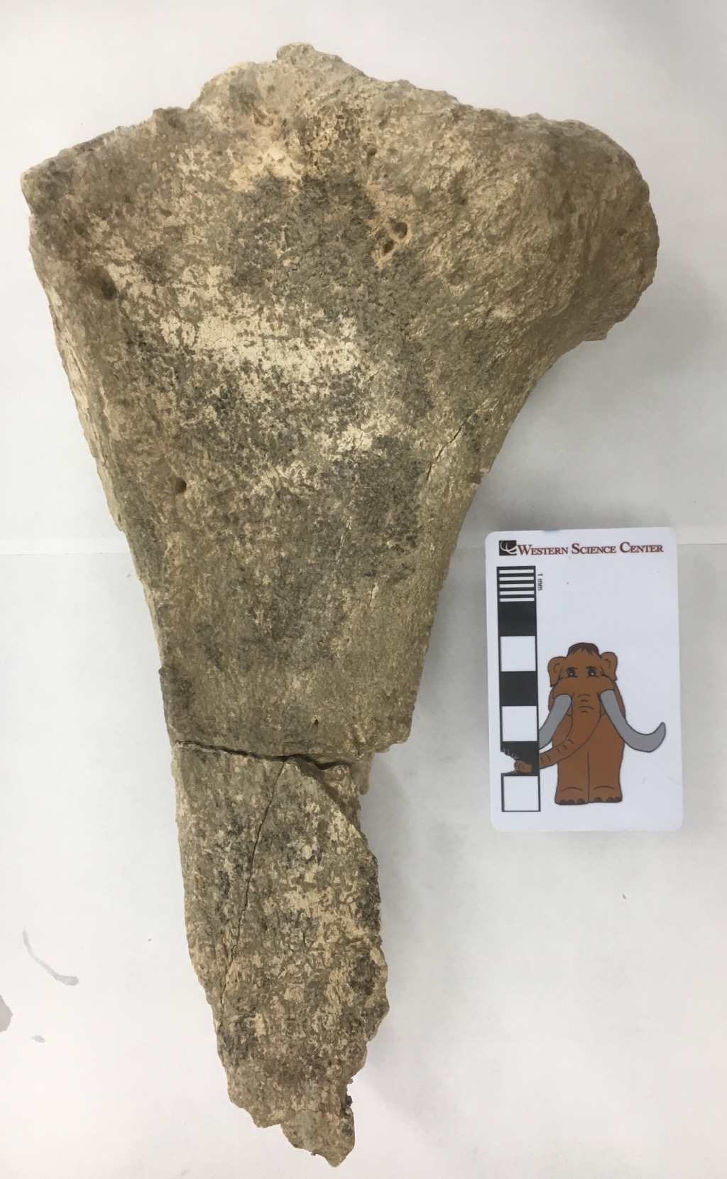 Over the last few weeks we've started pulling a lot of mastodon material from the collections (more on that in a future post). Some of the bones that are turning up are pretty interesting.The large bone fragment shown above is a small part of the proximal end of the right ulna, one of the bones in the forearm. It's shown above in lateral view, and below is looking at the proximal end (part of the articular surface for the elbow):
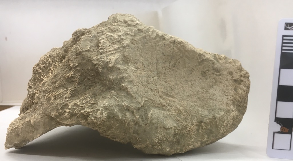 This fragment is labeled at mastodon, but comparing it to the photos in Olsen (1972) it seems to be closer to a mammoth (below). We'll have to examine it more closely and see if there's any associated material to determine for sure which taxon it belongs to
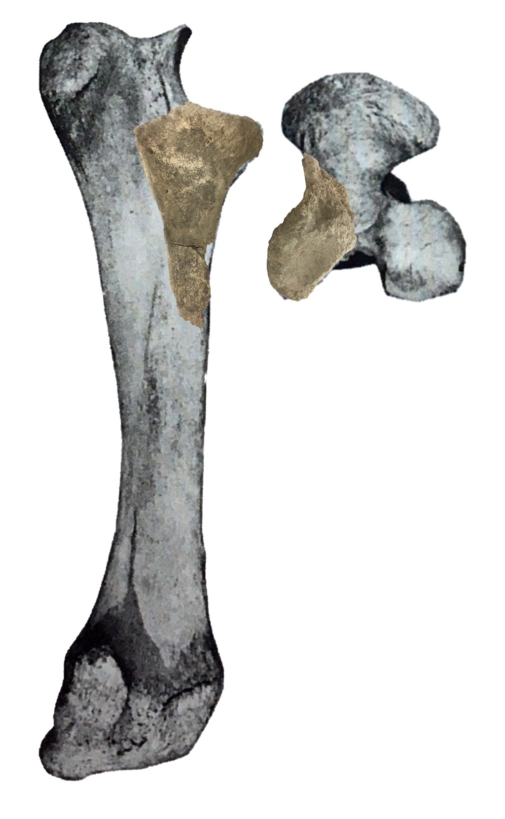 What really caught my attention were details on the edges of the articular surface and a few other places on the bone:
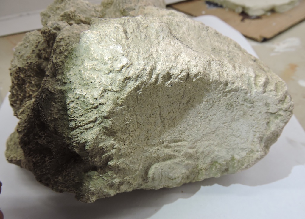
 As we're finding with many of the large bones from Diamond Valley Lake, this bone is covered with bite marks from scavengers, in the form of notches cut into the edges of the bone. These are relatively large grooves, consistent in size with something like a coyote or dire wolf, but there are lots of possibilities.
Reference:Olsen, S. J., 1972. Osteology for the Archaeologist No 3: The American mastodon and Wooly mammoth. Papers of the Peabody Museum of Archaeology and Ethnology, Harvard University 58:1-43.
Fossil Friday - chewed-up Bison tibia
 I recently finished reading Anthony Martin's book about dinosaur trace fossils, Dinosaurs Without Bones, so I've had trace fossils on my mind. Even though I'm not a trace fossil specialist I find them intriguing, because they are essentially fossilized behavior. The bone shown here is the distal part of a right tibia from a bison (it's from the older end of the valley, so it could be either Bison antiquus or Bison latifrons). Above is the anterior view, with the bottom of the bone (at the ankle joint) on the left. The proximal part, including the knee joint, is missing. Below is the posterior view of the same bone:
 Even in these views, you may have noticed that the distal end of the bone is a little misshapen. Close-ups reveal that this bone is absolutely riddled with bite marks, presumably from a predator/scavenger gnawing on the bone:


 The broken proximal end also has plenty of bite marks:
 This bone is crying out for a more detailed study. There appear to be at least 2-3 different sets of scratches with different widths. Does this indicate that there were different-sized scavengers? If so, are we looking at different species taking turns (maybe dire wolves followed by coyotes), or different ages of the same species (adult wolves and their pups)? If the scratches show cross-cutting relationships, it might be possible to figure out if the big animals were eating before the small ones, or the other way around, or at the same time.
Fossil Friday - camel lumbar vertebra
 While we only have one well-preserved skull of the extinct camel Camelops hesternus from Diamond Valley Lake, we have a large number of post-cranial remains.The bone shown above is a lumbar vertebra, seen in anterior view. This seems to be the 7th lumbar, the last one in the series before the sacrum (which was also recovered from this individual, along with several other bones). The prominent curved structures above and on each side of the neural canal are the prezygopophyses. These articulated with the postzygophyses of the 6th lumbar. The strong curvature would largely lock the two vertebrae together, resulting in a relatively inflexible lumbar region.Here is the posterior view, with the postzygophyses visible:
 And the left lateral view:
 The neural spine is broken on this specimen, as are the transverse processes (the right one is missing entirely). The broken surfaces are packed with sediment and abraded, indicating that they were broken off before burial. That is interesting considering that there are multiple associated bones with this specimen; that makes it less likely that the bone was damaged by, say, washing down a river. Could this damage have been caused by scavenging? I tried looking at the bone with low-angle light, which sometimes helps reveal bite marks on the surface:
 Sure enough, these are apparent bite marks on the bottom edge of the centrum. Below is the preserved part of the left transverse process, with apparent bits marks along its entire length:
 There are other possible bite marks scattered across this vertebra. Moreover, close examination also revealed possible insect feeding traces in various places, including on one of the prezygapophyses:
 These possible traces seem to be extremely common on bones from Diamond Valley Lake, possibly occurring on half or more of the large specimens. Clearly, besides studying the bones themselves, there's a lot of potential in the DVL trace fossils as well.
Fossil Friday - Carnivore traces
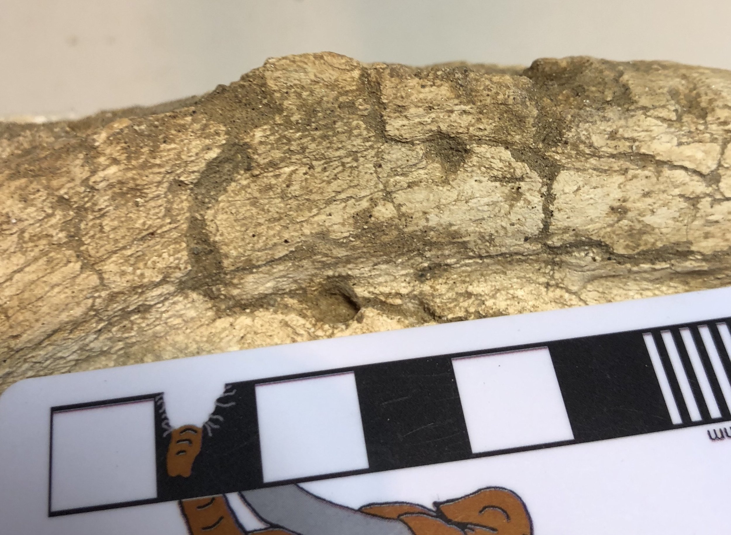
 In any large collection of vertebrate fossils, one of the more common specimen labels will be "unidentified bone fragment". But even an unidentified fragment can provide useful information. The bone shown above, found near the East Dam of DVL, has the following, rather uninspiring label: "Mammalia (larger size), unidentified bone fragment". This might be part of a transverse process or neural spine from a vertebra, and if so is could be from anything from a horse to a juvenile proboscidean. But there are also other possibilities; about the only things we can rule out are animals like rodents and rabbits that don't have any bones at all that are this large.So why is this bone interesting? Note the grooves visible in several places along the margin. Here are some views from different angles:
 These nearly-parallel grooves are consistent with gnaw marks from a carnivoran, so this fragment is covered with trace fossils. We can't say with certainty what type of carnivoran made the marks, but they are not particularly large, so it's unlikely we're looking at a huge animal like Arctodus. A medium-sized carnivoran such as a black bear, dire wolf, or coyote is a more likely possibility, or perhaps even a badger; all of these are known from the Diamond Valley Lake deposits.As I've mentioned in previous posts, carnivoran bones are quite rare in the DVL deposits. But we do see evidence of their activity. We haven't yet catalogued all the bite marks that are present on DVL bones, but I suspect we actually have more carnivoran trace fossils than bones in our collection.
Fossil Friday - Stories from Bones exhibit
![]() For Fossil Friday this week, I want to highlight Western Science Center's new exhibit "Stories from Bones", which opens tomorrow.While WSC has excellent paleontology exhibits, as with any museum with a large collection many of the specimens are not on public display. There are a variety of reasons for this. Of course, the biggest obstacle is money; cases, information panels, interactive, floor space, and other requirements for an effective display are all expensive, and even the healthiest museums operate on a shoestring budget. Besides money issues, many specimens are just not suitable for display. Perhaps they're too fragile to risk moving around too much, or too fragmentary to interpret for the public (a specimen that visually looks like a piece of junk can still produce valuable scientific data). Even with all these limitations, we strive to make as much of our collections accessible to the public as possible. "Stories from Bones" is a result of that effort.
For Fossil Friday this week, I want to highlight Western Science Center's new exhibit "Stories from Bones", which opens tomorrow.While WSC has excellent paleontology exhibits, as with any museum with a large collection many of the specimens are not on public display. There are a variety of reasons for this. Of course, the biggest obstacle is money; cases, information panels, interactive, floor space, and other requirements for an effective display are all expensive, and even the healthiest museums operate on a shoestring budget. Besides money issues, many specimens are just not suitable for display. Perhaps they're too fragile to risk moving around too much, or too fragmentary to interpret for the public (a specimen that visually looks like a piece of junk can still produce valuable scientific data). Even with all these limitations, we strive to make as much of our collections accessible to the public as possible. "Stories from Bones" is a result of that effort.
 Mammoth jaw display in "Stories from Bones".
An important aspect of planning an effective exhibit is developing a theme. An exhibit is telling a story, and you need to be aware of what that story is as the exhibit is being designed. The theme might be "We have a bunch of stuff!", but while that was a common theme in museums a century ago (and one I personally appreciate), it does not generally make for the most informative exhibit experience for the majority of visitors.Once the theme is established, it's important to stick to it, so that the exhibit story remains coherent. Imagine reading a mystery novel in which three chapters are devoted to a history of the development of the gunpowder used in the crime, and an additional chapter describes the etymology of the last name of the victim, when neither is important to the outcome of the story. Each of these things might be individually interesting, but if you try to talk about all of them then you risk obscuring everything. There is a real risk of this "mission creep" in an exhibit based on a data-rich field such as paleontology. We might talk about evolutionary relationships, paleoenvironmental indicators, biogeographic information, site-specific descriptions, or an array of other things. Talking about any of these might be a good idea; talking about all of them is a bad idea.The permanent paleontology exhibit at WSC does this very well. The exhibit is basically a review of the Diamond Valley Lake Local Fauna; what was here, how does it compare to the rest of Southern California, and (as a secondary point) what does it tell us about the local Pleistocene paleoenvironment. In contrast, "Stories from Bones" asks "What do these fossils tell us about the lives and deaths of these individual animals?".To that end, "Stories" has a series of displays that talk about how paleontologists determine how old an animal was when it died. We have several cases that look at tooth replacement in proboscideans, horses, and bison, such as the two mammoth jaws above (they're close to the same size, but one animal was about 30 years older than the other), or the three bison dentaries shown below that represent young, middle-aged, and elderly animals.
 Bison jaw display in "Stories from Bones".
We have several examples of bones that were broken and healed, evidence of events that took place during an animal's life:
 Broken and healed bones in "Stories from Bones".
We also have several cases that describe taphonomic features, looking at what happened to an animal at or immediately after death.We designed and built a number of interactive displays for this exhibit. The most prominent is a cast and video of the CT scans of Max the Mastodon's lower jaw, taken back in August.
 Max's CT-scan station in "Stories from Bones" during installation, under the watchful eye of @MaxMastodon.
We're proud of the fact that several of our interactive displays ask visitors to map or measure specimens and reach conclusions based on their data:
 A more extensive version of the bison tooth display shown here is also available as a guided activity for school groups visiting the museum, and as a kit available for purchase.If you're a regular reader of this blog, you'll find that "Stories from Bones" draws heavily from my past "Fossil Friday" posts. For most of those specimens, this is the first time they've ever been on public display, so if you're near Southern California make sure to stop by the museum. "Stories from Bones" opens on October 31, and will remain open into May 2016.
Western Science Center theropod invasion?
 I arrived at work this morning to find what appeared to be several muddy tracks in the museum parking lot. While they weren't arranged in an organized trackway, they were numerous.
  These look a lot like small theropod dinosaur tracks, complete with claw impressions, such as this one from Dinosaur State Park in Connecticut:
 Of course, my first thought was that the museum was under attack from a pack of angry, ravenous theropod dinosaurs, presumably seeking revenge for my disparaging comments about the Jurassic World "Velociraptors". Upon further reflection and observation, it turned out that there was a more mundane explanation. The museum has several sweetgum trees around the parking lot. These leaves are generally five-pointed (below), but when the leaves fall and dry out in the sun the edges tend to curl up.
 We've actually had a little rain in Hemet the last few days, and the ground is a bit muddy. The leaves apparently got muddy on one side, then blew into the parking lot overnight. Then, before morning, the leaves blew away, leaving a three-point mud outline (the curled-up lateral portions of the leaves apparently didn't touch the ground).As a result, I was able to make it safely to my office, free of any Velociraptor-related unpleasantness.
Fossil Friday - "Then the rats got him"
 Bison are among the most common large animals in the Pleistocene Diamond Valley Lake fauna, but like almost all the remains from these deposits they are usually fragmentary. But even fragmentary fossils can provide a lot of information, including the bison right lower jaw fragment shown here.Sometimes bonebeds or other rich deposits of fossils are formed during catastrophic events, with large numbers of organisms buried together very rapidly. That's not what we see in Diamond and Domenigoni Valleys, however; these deposits are primarily the result of the gradual accumulation of skeletons over thousands of years. That means that an individual skeleton may have laid exposed at the surface for a significant period of time before it was eventually buried. This exposes the bones to sun, wind, and rain, all of which can cause the bone to deteriorate and the skeleton to fall apart, and to scavengers, which may damage or eat the bones, or carry pieces away. All these things contribute to the fragmentary nature of the Diamond Valley Lake specimens.Evidence of the specific types postmortem trauma experienced by these bones is sometimes preserved. If we zoom in on the bison jaw shown above, we can see a series of grooves cut into the bone:
 These grooves are bite marks, most likely made by rodents. Bone gnawing has been documented in lots of different rodents, including rats, mice, and squirrels. It's generally attributed to the need for rodents to keep their ever-growing incisors from becoming too long, and perhaps to provide an additional source of calcium. But the presence of these marks immediately tells us quite a bit of additional information; the bison died in a setting that didn't result in immediate burial, and rodents were present in the environment.Of course, from the bison's point of view, maybe this is only adding insult to injury!
Fossil Friday - bite marks on a camel skull
 One of the specimens we have on display at the Western Science Center is a cranium and partial vertebral column including the neck of the camel Camelops hesternus. A closer examination of the skull reveals some surprising features. The parietals (the bones that make up the back half of the top of the braincase) have a series of holes and apparent scrapes:
 At first I thought the two back holes might be an anatomical feature called the parietal foramen. However, parietal foramina are uncommon, usually forming as a developmental abnormality. I've been unable to find images or reports of parietal foramina in Camelops or any other camel. The holes in this specimen are not symmetrical in their position (each one is located in a different position on its respective parietal), and there are cracks in the parietals leading to the holes. Finally, if these were parietal foramina, it doesn't help to explain the presence of the other hole located at the front of the parietal. Taken together, this suggests that the holes are not an anatomical feature but instead are bite marks from another animal.I originally envisioned a carnivore grabbing the camel by the top of the head, with its head held almost parallel to the camel's head, and then dragging the camel's head and neck away from the rest of the carcass. This would mean that the four parietal marks (three punctures and one scrape) were made by the four canines of the carnivore in a single bite. Darla and I pulled out cast skulls of several different carnivores to try this out, but we couldn't get it to work. If we assume that the four marks were made in a single bite, then the spacing is about right for a small dire wolf, a black bear, or a jaguar. However, when we tried to simulate the bite the predator's incisors would always hit the sagittal crest (the ridge of bone along the top of the skull) long before the canines reached the parietals. If we went with larger animals with longer canines like a short-faced bear or an American lion, the canines would reach the parietals but the spacing was far to close for such a large animal.I now think that it's more likely that the punctures were caused by multiple bites, by a carnivore coming at the skull from an oblique angle (possibly multiple angles), something like this:
 This actually may be more consistent with the behavior of modern carnivores; if you want gory confirmation, do an image search for "hyena carrying head" for examples of how scavenging carnivores transport prey. There is also some evidence that there may be additional bite marks all over this skull, consistent with multiple bites. There are two unusual depressions in the top of the frontal bone, several holes along the edge of the right lambdoidal crest at the back of the skull, and damage to part of the right squamosal. To be fair, none of these additional marks are sure things; if not for the four bite marks on the parietal I would have never considered these as likely candidates for bite marks.Another observation I would not have thought twice about except for this context is the damage at the front of the skull. The nasal bones are damaged, and the premaxillary bones are missing entirely. The premaxillae can loosen and fall off as a skull dries out, and that's normally how I would explain this. But if this skull was being chewed up by carnivores it raises another possibility. It seems that the nose is one of the choice bits of meat on the skull for a carnivore. There's a lot of meat and blood in the nose, and it's easier to get to than the brain. Moreover, while cats such as lions usually kill their prey with a bite to the neck, closing the windpipe, an alternate method used on occasion is to bite down on the nose. Is it possible that the missing premaxillae in this specimen is another predation feature?