

 Today's Fossil Friday specimen comes from the Pleistocene camel Camelops hesternus, a taxon we've featured several times on this blog. But this specimen is special because of where it was found - in Joshua Tree National Park.Joshua Tree NP is best known for its eponymous trees and spectacular granite exposures, but the park also preserves delicate desert habitats, native cultural artifacts, and fossils. A few months ago Western Science Center became a repository for Joshua Tree, and dozens of vertebrate fossils collected under the direction of Kathleen Springer were transferred here. One of the specimens is the tooth shown here, a lower right third molar from Camelops.To the extent that dating these fossils has been possible, they are roughly equivalent in age to the Diamond Valley Lake and Rancho La Brea fossils. Yet there appear to be real faunal differences distinguishing Joshua Tree from other localities, and there is a lot of exciting research ahead for these fossils.Another thing in their future is an exhibit. WSC is opening a new temporary exhibit, "Fossils of Joshua Tree National Park", which will include representative fossils from this collection. There is an opening reception for museum members tonight, and the exhibit will open to the general public tomorrow morning. Thanks to Vincent Santucci from the National Park Service, Melanie Spoo and the staff of Joshua Tree National Park, and Kathleen Springer of the USGS for making WSC the fossil repository for Joshua Tree and making this exhibit possible.
Fossil Friday - Miocene horses
Hello! I'm Brittney Stoneburg, the Marketing and Events Specialist for the Western Science Center. While my job mostly entails communications and outreach at the museum, I've spent the last year dipping my toes into research!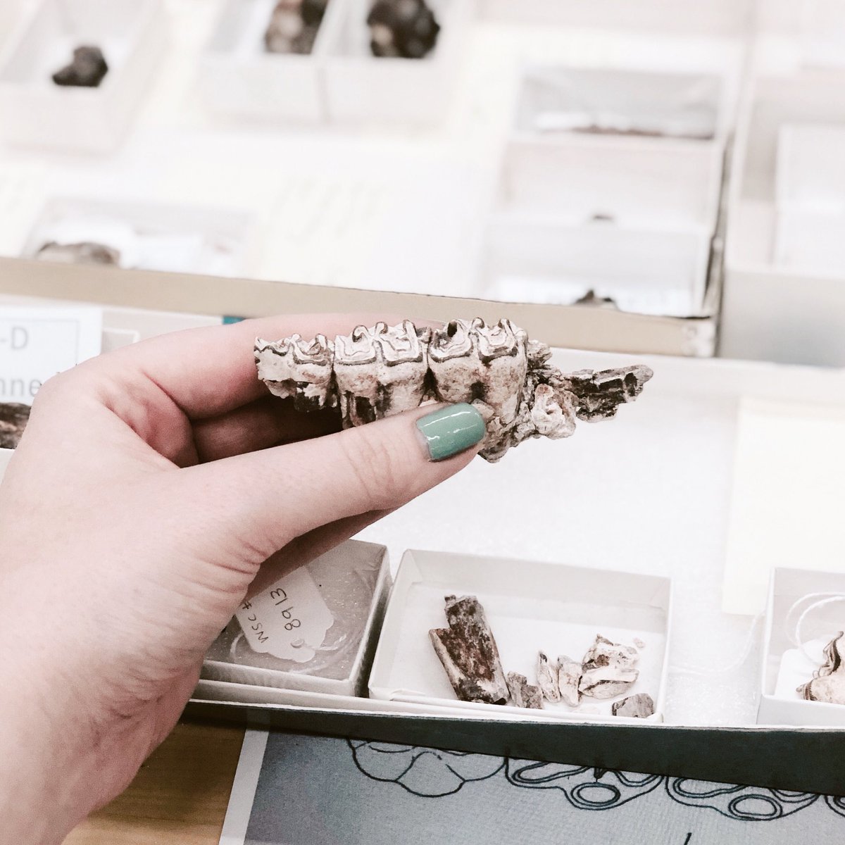 For my first research project, I've been working on fossils collected by the Western Science Center from our field site in San Bernardino National Forest. This means I'm up to my neck in horse teeth right now! I've been focusing on the small, three toed horses that roamed Southern California during the early to middle Miocene, approximately 18 million years ago.
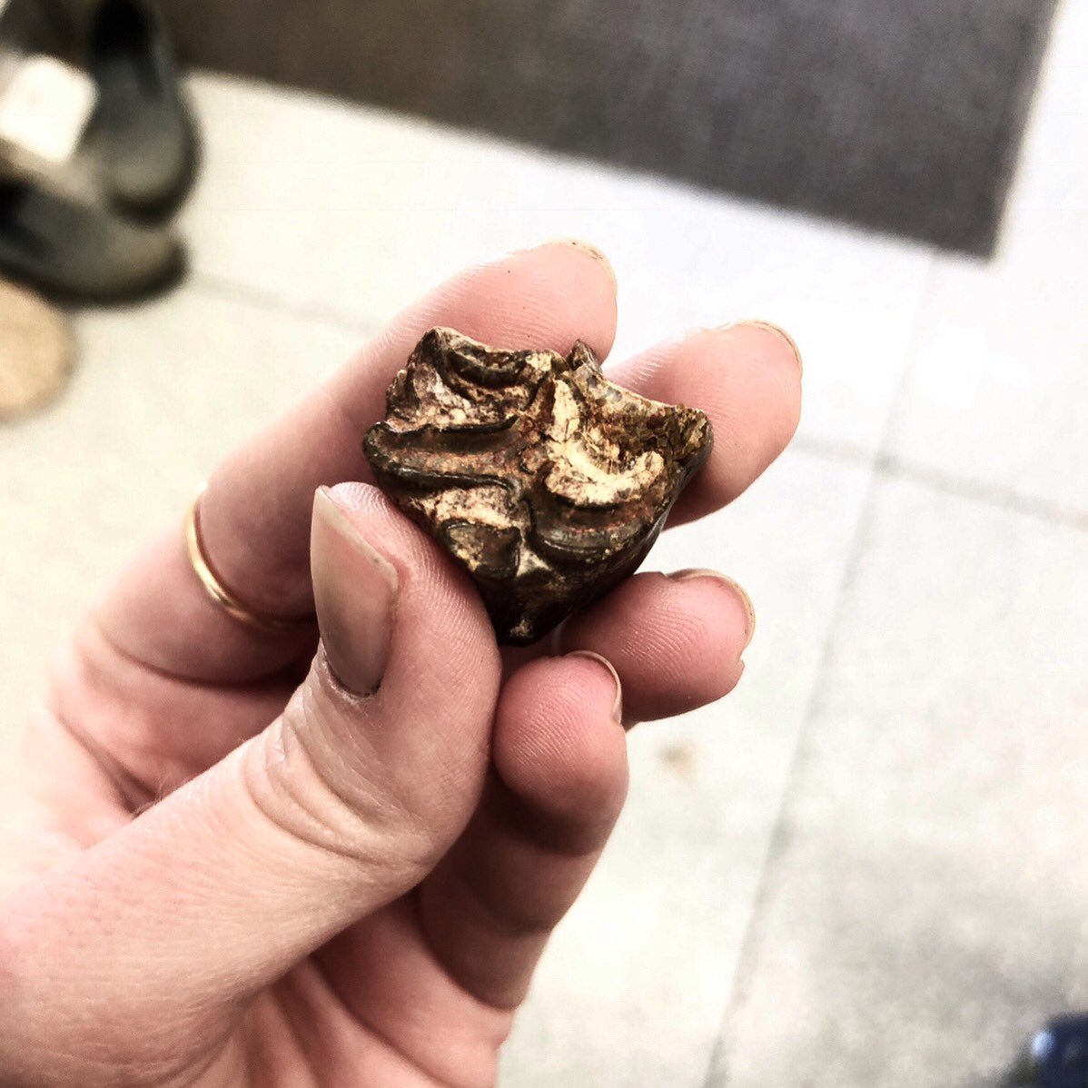 Multiple species of horse lived in this area, including Scaphohippus, whose teeth are picture above. While our collection from this site has only a few post-cranial bits, we can make pretty solid identifications just from the teeth. Horse teeth like these have numerous features that make it possible to identify them down to the species level. Knowing what horses lived in this area during the Miocene can tell us a lot not only about the horses themselves, but about the environment they lived in!I'll be presenting on my research at the North American Paleontological Conference in June, so if you'll be there, feel free to ask me about all of these horses!
Fossil Friday - ceratopsid illium
 Last week for Fossil Friday, I posted the sacrum of a ceratopsid, a large horned dinosaur related to Triceratops. The sacrum is a series of fused vertebrae to which the hip bones attach. Today, I want to show you one of those hip bones from the same ceratopsid individual. This is the left ilium, showing the inner surface where it would have attached to the sacral ribs along a series of deep facets. At about 80 centimeters long, this ilium is not terribly big for a ceratopsid, but it still would have been a hefty animal, plodding around the forests and swamps of Late Cretaceous New Mexico.In the coming months, we will create 3D-printed replicas of the sacrum and ilium of this ceratopsid to see how they fit together, the first step in rebuilding this 79-million-year-old skeleton. We also have several vertebrae and ribs from the back. This specimen was collected during the 2017 and 2018 field seasons by staff and volunteers from the Western Science Center, Zuni Dinosaur Institute for Geosciences, and Southwest Paleontological Society. The sacrum and ilium have both been prepped by WSC lab volunteer Joe Reavis.Post by Curator Dr. Andrew McDonald.
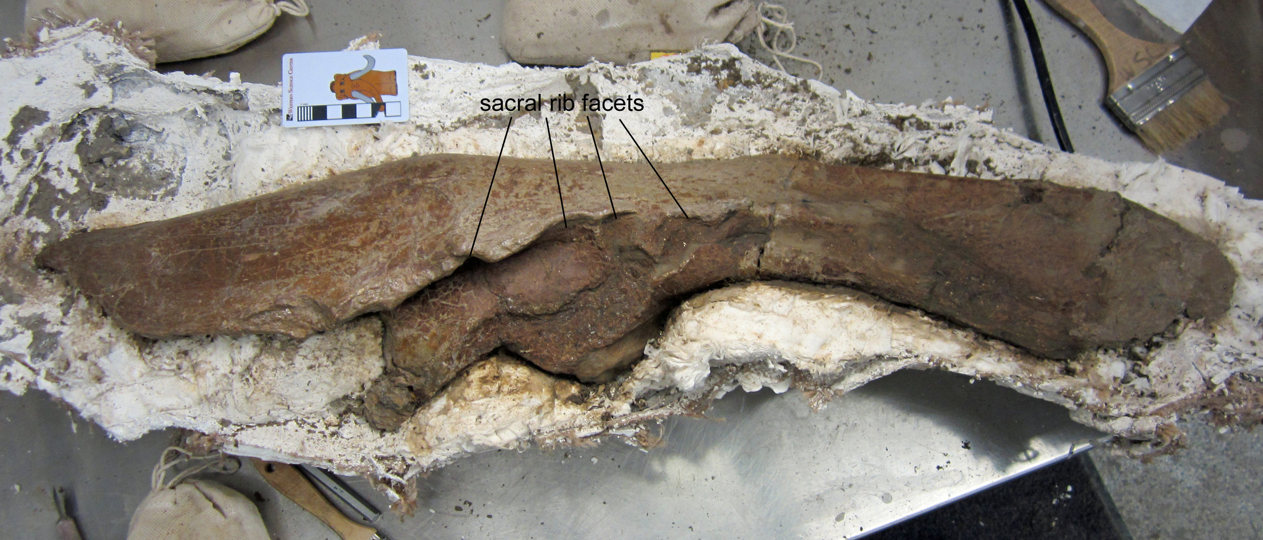
Fossil Friday - ceratopsian sacrum
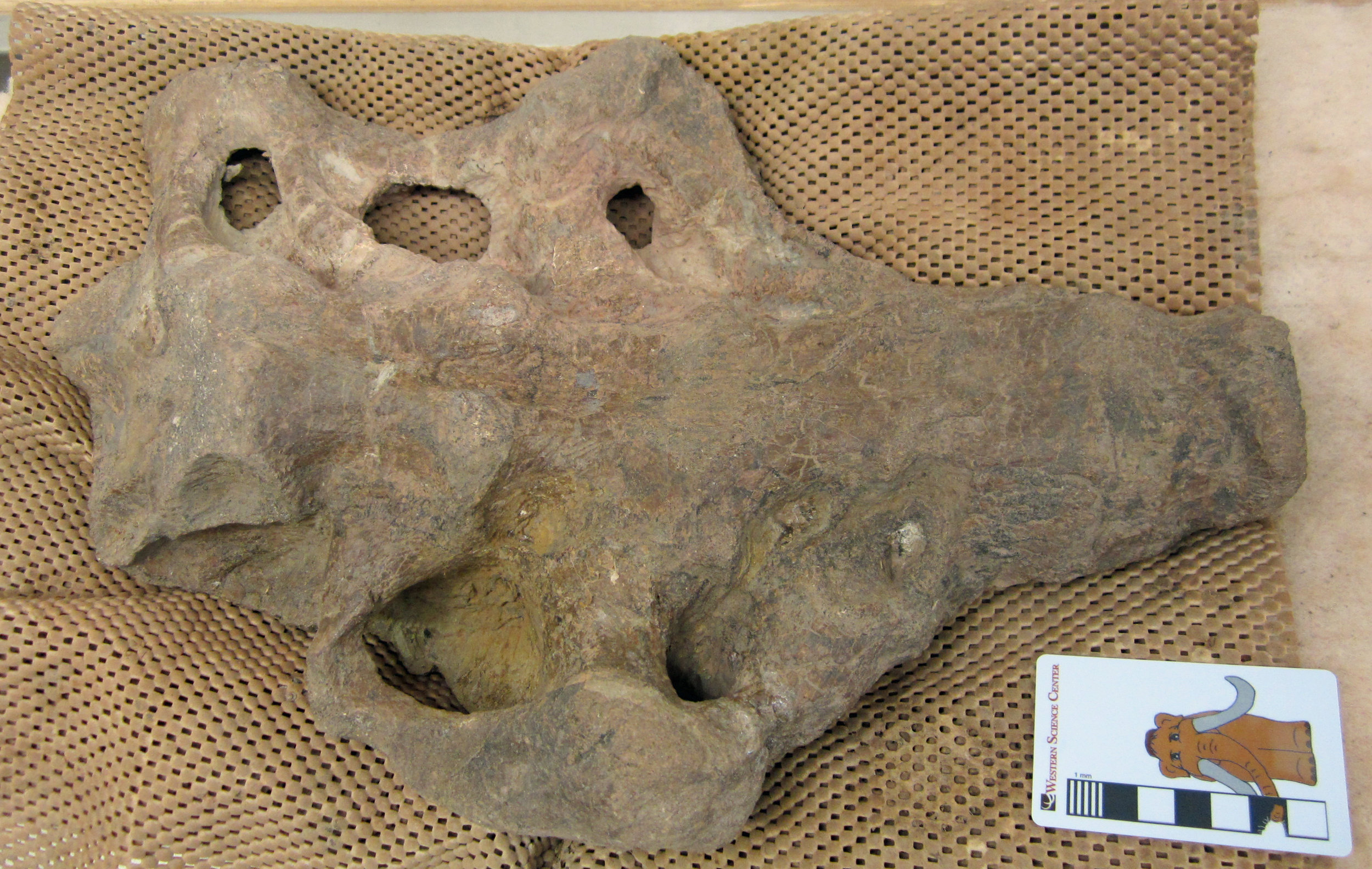 Horned dinosaurs were one of the most successful groups of dinosaurs in the Late Cretaceous of western North America. Known as ceratopsids, these rhino- to elephant-sized beasts brandished horns, spikes, and frills on their massive skulls. More than 40 different species are known to have inhabited western North America at different points in time between 80 and 66 million years ago. The last of them was the great "three-horned face" - Triceratops.Ceratopsids have been known from the Menefee Formation of New Mexico since the 1990s, when paleontologists at the New Mexico Museum of Natural History & Science reported fossils including a partial skeleton. More recent expeditions to the Menefee by the Western Science Center, Zuni Dinosaur Institute for Geosciences, and Southwest Paleontological Society have discovered further ceratopsid fossils, including another partial skeleton now being prepped at WSC.Today's image is the sacrum of that 79-million-year-old ceratopsid skeleton, recently prepped by WSC volunteer Joe Reavis. The sacrum is a series of fused vertebrae to which the hips were attached. In the image, the bottom surface of the bone is visible. We'll be working on more bones from this skeleton over the coming months, including a hip bone, more vertebrae, and ribs.Post by Curator Dr. Andrew McDonald.
Fossil Friday - fern
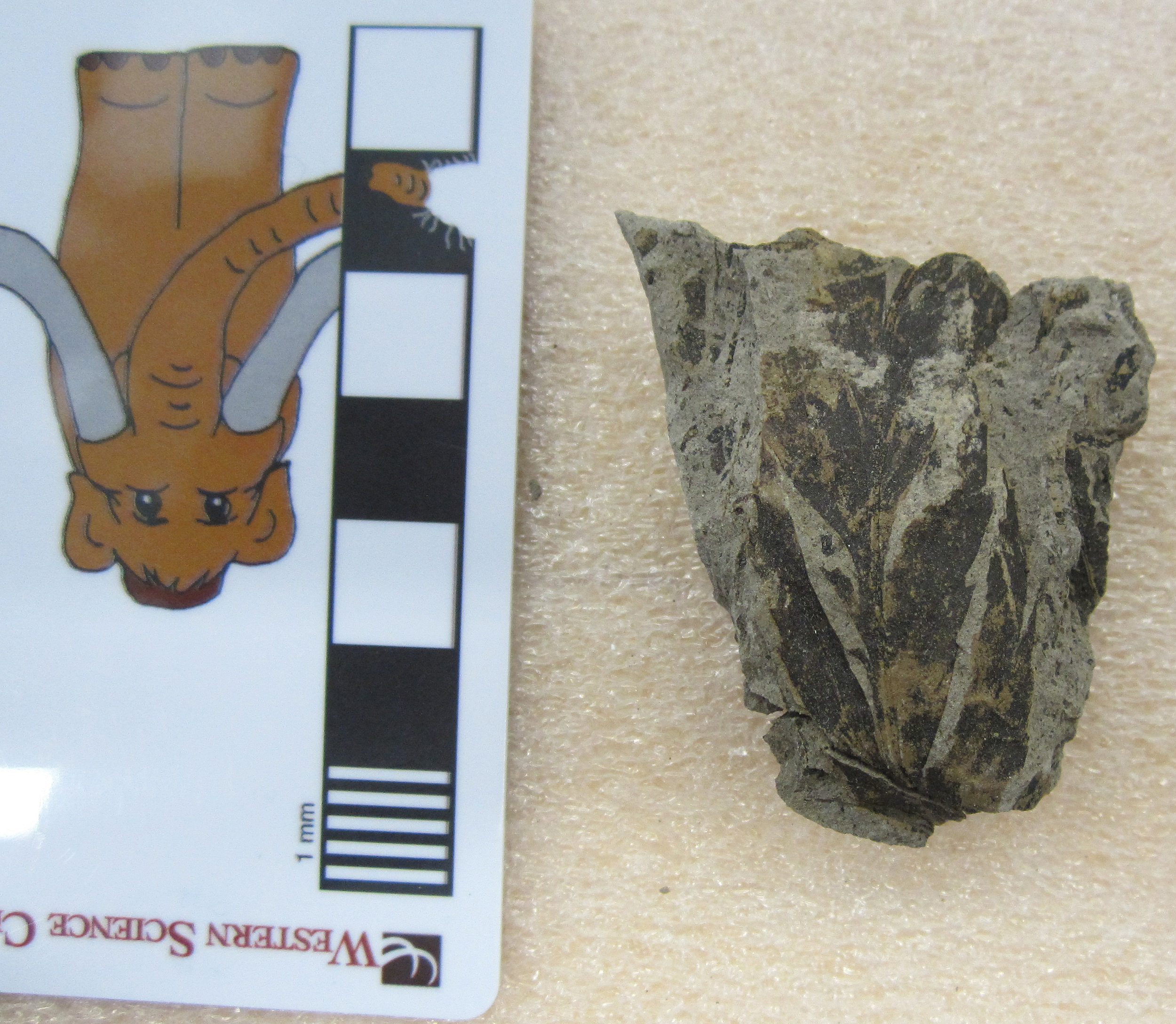 Over the last year or so, I've posted many bones from large herbivorous dinosaurs that lived in New Mexico around 79 million years ago, such as duck-billed hadrosaurs, horned ceratopsids, and the armored Invictarx.Alongside the bones of these beasts in the Menefee Formation we often find fossil plants. Many of these occurrences are petrified logs and stumps, which we find eroding out onto the surface.However, sometimes when we dig into the layers of mudstone to excavate dinosaur bones, we also encounter beautiful fossil leaves. The image today is a very nice leaf from a Late Cretaceous fern, probably belonging to the living fern genus Anemia. Fossils like this give us some idea of what the plant-eating dinosaurs might have been eating, and show that back then New Mexico was a much wetter habitat than today. It was a muddy floodplain lush with plants and teeming with dinosaurs.Post by Curator Dr. Andrew McDonald
Fossil Friday - Mistaken Identity
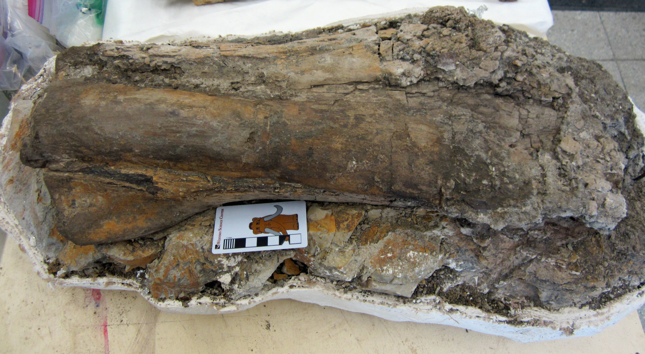
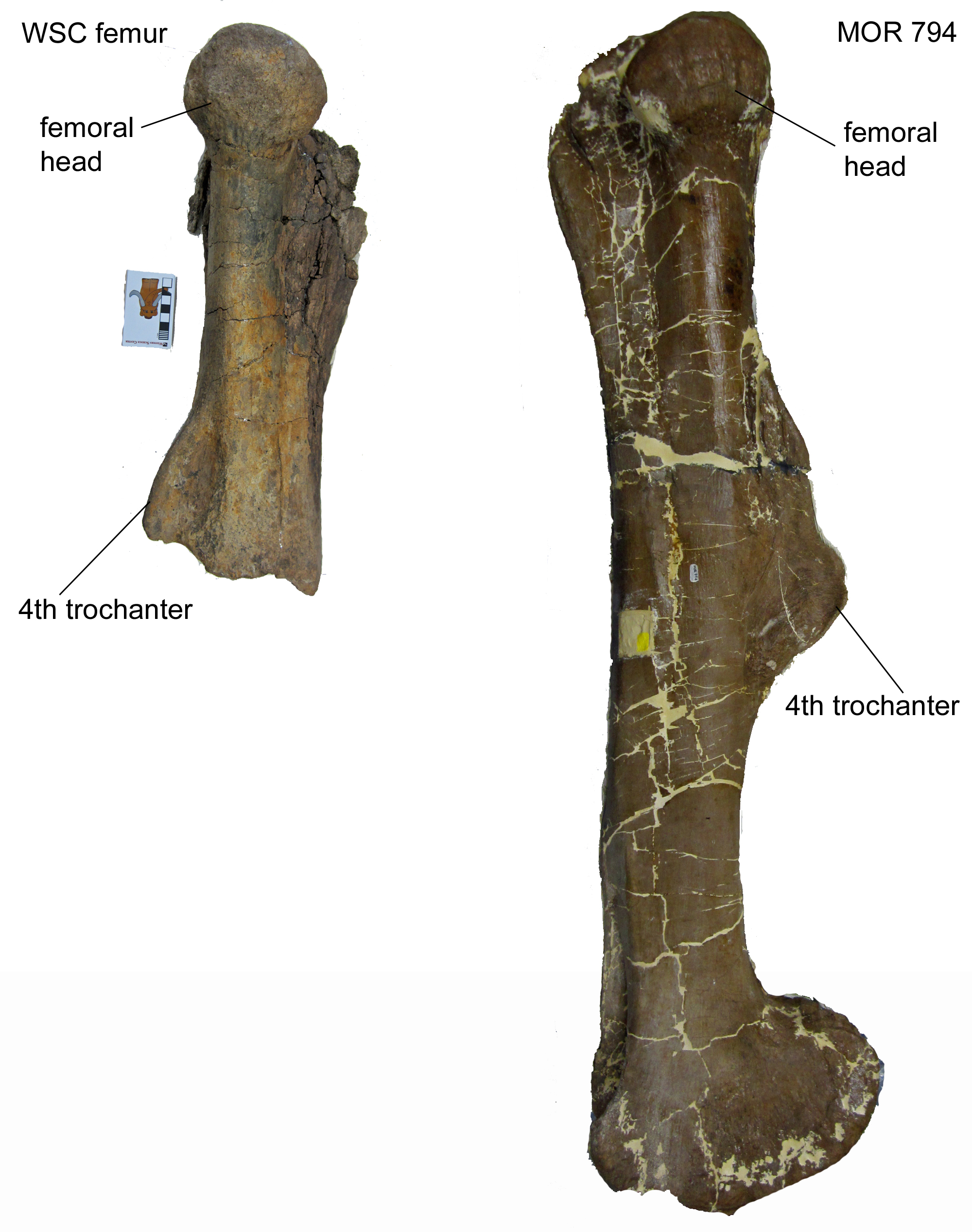 On March 8 Fossil Friday, I posted an 80-million-year-old dinosaur bone from New Mexico, which I identified as a tibia (shin bone). At the time, it was only partially prepped, and since then, WSC volunteer Joe Reavis has been working tirelessly to remove the remaining mudstone.It turns out the "tibia" is actually a femur! It's the proximal half of the left femur of a hadrosaur, one of the large duck-billed plant-eating dinosaurs that are so abundant in Upper Cretaceous rocks throughout the American West. Comparison with a complete hadrosaur femur from Montana (Museum of the Rockies specimen MOR 794) allows us to identify several major features, including the head that fitted into the hip socket and the 4th trochanter, a major muscle attachment site. It also shows that the WSC femur is from a fairly large individual, probably close to 30 feet long.Hadrosaur bones are very common in the Menefee Formation, the rock layer in which this femur was collected by my colleagues from the Zuni Dinosaur Institute for Geosciences and Southwest Paleontological Society in 2015. We'll have more to say in the near future.Post by Curator Dr. Andrew McDonald
Fossil Friday - Paralia sulcata
 "Max's Minions" is the informal name for WSC's junior research volunteer program. The Minions perform a variety of lab duties for us, including 3D scanning, molding and casting, skinning carcasses for our dermestid colony, preparing fossils, and other tasks. Several of them are also working on their own research projects.Western Center Academy student Sianna Xiao has been pulling sediment samples off our small collection of fossil bones and shells from Neogene deposits in Virginia. Using a protocol developed some years ago by one of my former student interns, Anna Trochim, she has been preparing those samples for studying their diatoms.Diatoms are single-celled eukaryotic algae that live in the water. An important characteristic of diatoms for paleontologists is that they secrete a cell wall (called a frustule) that is made of silicon dioxide; essentially, they have glass shells. This means that they preserve well as fossils.Diatoms are one of the main things we use to determine environmental conditions, as various species may be sensitive to changes in salinity, temperature, pH, or other factors. Since they are photosynthetic, they depend on sunlight, so forms that live on the seafloor can only live in shallow water (less than 100 m, and usually less than 20 m). They are also one of the main organisms used in Cenozoic marine biostratigraphy, meaning they are important for determining the age of marine deposits. So there is an abundance of reasons to study diatoms.One of Sianna's samples comes from the Miocene Calvert Formation at the Carmel Church Quarry. Anna and I published the Carmel Church diatom flora a few years ago, but since we know it contains diatoms it's a good way to learn the sample protocol and practice identifications. The image above is one of the most common species at Carmel Church, Paralia sulcata. As expected, it's one of the most common in Sianna's slides as well. This species, while common, has a tremendous temporal range m(from the Cretaceous to Recent), so it's of no biostratigraphic value. It also can live in seawater ranging from brackish to normal salinity, and also lives in a variety of environments, so it doesn't actually tell us a whole lot about the age or paleoenvironment. But 27 other species are known from Carmel Church so far, so there will be plenty more to see.
Fossil Friday - abelone
 Last week Cogstone Resource Management (http://www.cogstone.com) delivered a collection of Pleistocene deposits from Ventura County to the Western Science Center. This included a number of both vertebrate and invertebrate fossils.These photos show a shell from an abelone, Haliotis sp.. While the shell looks a lot like a clam, abelone are actually snails; the light-colored early whirls of the shell are visible adjacent to the scale bar. It's also clear on the other side of the shell that it's not from a clam, as there is no hinge line to articulate with a second shell (below). This view also shows off the opalescence for which abelone are famous.
 Abelone typically live in fairly shallow water just below the intertidal zone, where they feed on algae. Associated fossils in this collection also indicate a similar shallow marine setting.This abelone and other fossils from this site will be featured in an exhibit opening at WSC this fall. Thanks to Cogstone for bringing it to us!
Fossil Friday - Mammut pacificus
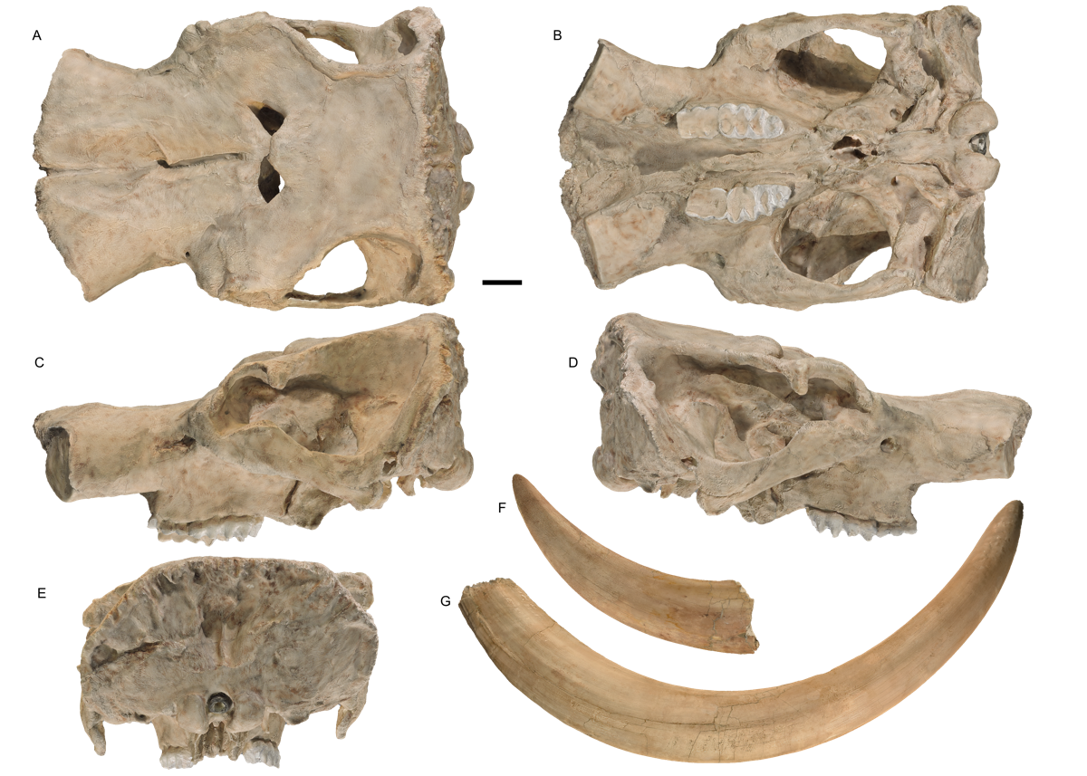 The big news this week for Western Science Center was the naming of a new species of mastodon, Mammut pacificus.For decades, the consensus on Pleistocene mastodons (which I shared) was that in North America there was only a single, widespread species, Mammut americanum. Four years ago, I stumbled across the fact that California mastodons have different tooth proportions than other mastodons. A group of us started exploring that issue, trying to determine what was going on. To that end, in 2016 we launched a crowdfunding campaign on experiment.com to allow us to gather mastodon measurements from other parts of the continent. Exploring this problem continued through the Valley of the Mastodons symposium in 2017. Sometime in late 2017, we started to consider the possibility that we were seeing these differences because we were dealing with two different species, and by mid-2018 we started working on a manuscript. It was finally published last Wednesday.So, what makes the Pacific mastodon different from the American mastodon? Here are the major characters:-- Mammut pacificus has narrower teeth than M. americanum, especially the 3rd molars. On the left below is a lower third molar from Max, and on the right is the same tooth from an American mastodon from Ohio (from the Cincinnati Museum of Natural History and Science).
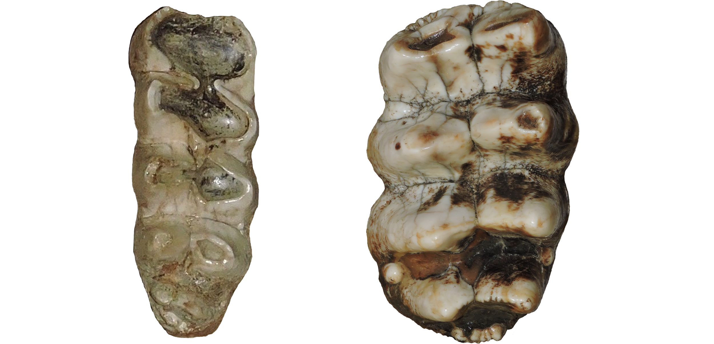 This trend is present in every tooth position except the upper 2nd molars, although we had the best statistical support for the 3rd molars.-- Mammut pacificus has six sacral vertebrae, while M. americanum usually has five. There has been one American mastodon reported with six sacrals, and another reported with four (although I have not personally seen either of these specimens), but all the other specimens for which we could get data (five of them) have five sacrals. All three known M. pacificus sacrals, including the one below from Diamond Valley Lake, have six.
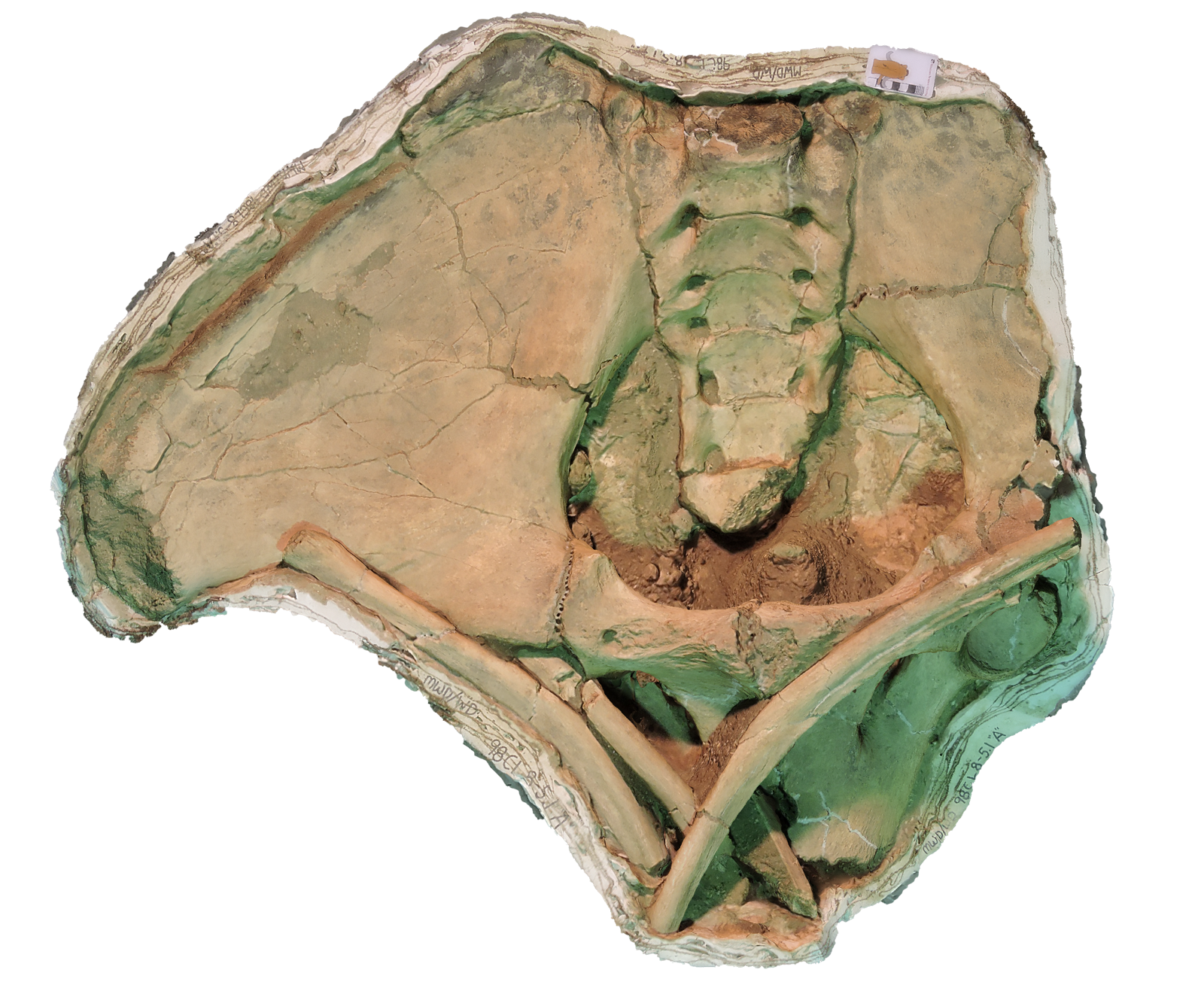 -- Mammut pacificus has a thicker femur for a given length. This is a feature we need to explore further, but our data is a bit limited. The trends we see suggest that not only do Pacific mastodons have thicker femora, but that the difference becomes more pronounced with larger body size. However, we don't have any really big M. pacificus femora to confirm this (Max's femur is incomplete). Below, "Little Stevie's" femur from DVL is on the left, compared to an American mastodon specimen from Ohio (again, from the Cincinnati Museum).
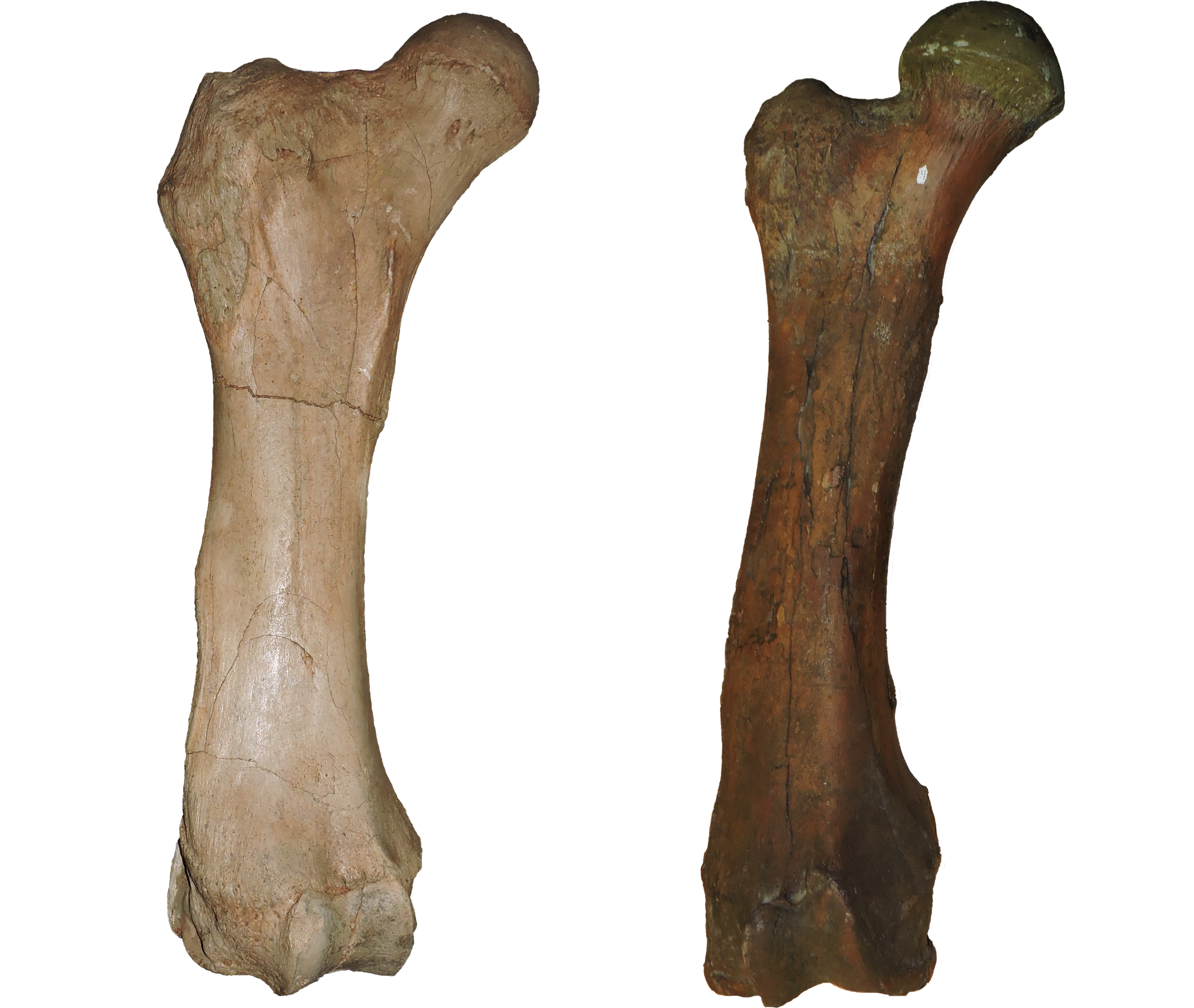 -- Mammut pacificus has no mandibular tusks, while they still occurred in about 25% of the M. americanum population. This is a tricky character, because both taxa seem to have been losing their chin tusks (the presence of chin tusks is the primitive condition for mastodons). But it seems that Pacific mastodons got there first. Below, at the top is Max's lower jaw, and below is a lower jaw from Colorado (from the Denver Museum of Nature and Science).Below is a map from our paper showing all the known Mammut pacificus localities, as well as the Mammut americanum localities we used in this study (there are many, many additional American mastodon sites we didn't examine):
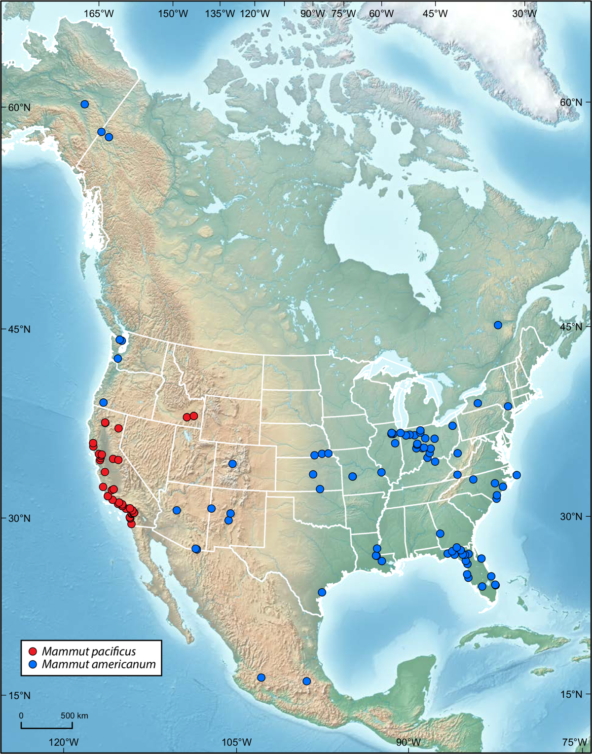 Note that, with the Oregon sample, we did not find any of the diagnostic bones (m3, femur, sacrum, lower jaw), so we conservatively coded the data as M. americanum, but it could actually be M. pacificus.We are committed to making our discovery as widely known as possible. To that end, we published our paper in the open access journal PeerJ, where it is available as a free download. We have also made 3D scans of 24 of the key DVL specimens available on MorphoSource and Sketchfab.I'll be writing more about the Pacific mastodon, but this is a quick summary of the highlights of the paper. If you really want to get into the details, our data is all available at the PeerJ link. Enjoy!
Fossil Friday - mammoth molar
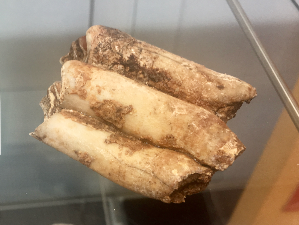
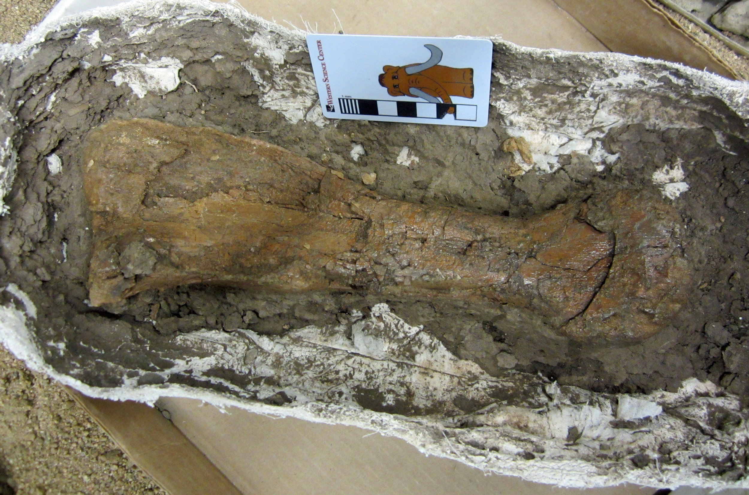
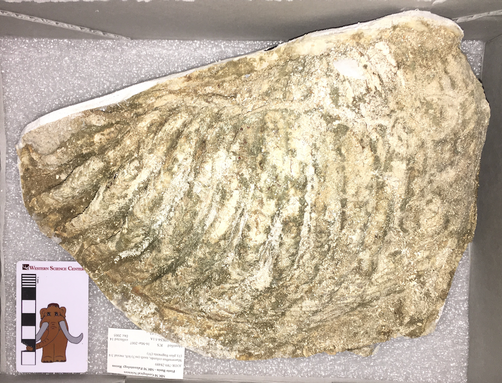 Today's Fossil Friday specimen comes from a location we've never before featured on this blog, Joshua Tree National Park.Joshua Tree is most famous for its eponymous trees and its spectacular granite formations, but there are also significant Pleistocene fossil deposits within the park. Over a period of several years Kathleen Springer and Eric Scott (then at the San Bernardino County Museum) collected some of these remains under a NPS permit. Earlier this week Joshua Tree designated Western Science Center as the repository for that collection and transferred the specimens here.The Joshua Tree fauna is an contrast with other Southern California sites such as Diamond Valley Lake in that, like today, during the Pleistocene it was more mountainous and more arid than the inland valleys. While the fossils from Joshua Tree are almost all fragmentary and poorly preserved, studying the fauna from this site will help give us a more complete understanding of how California ecosystems worked during the Pleistocene.The particular fossil shown here is a Columbian mammoth lower left third molar, one of several mammoth remains in the collection. Mammoths seem to be one taxon that had a great deal of ecological flexibility, as they are found in almost every imaginable Pleistocene deposit.Thanks to Melanie Spoo of Joshua Tree National Park and Vincent Santucci of the National Park Service for arranging the move of the Joshua Tree fossils to WSC.
Fossil Friday - dinosaur tibia
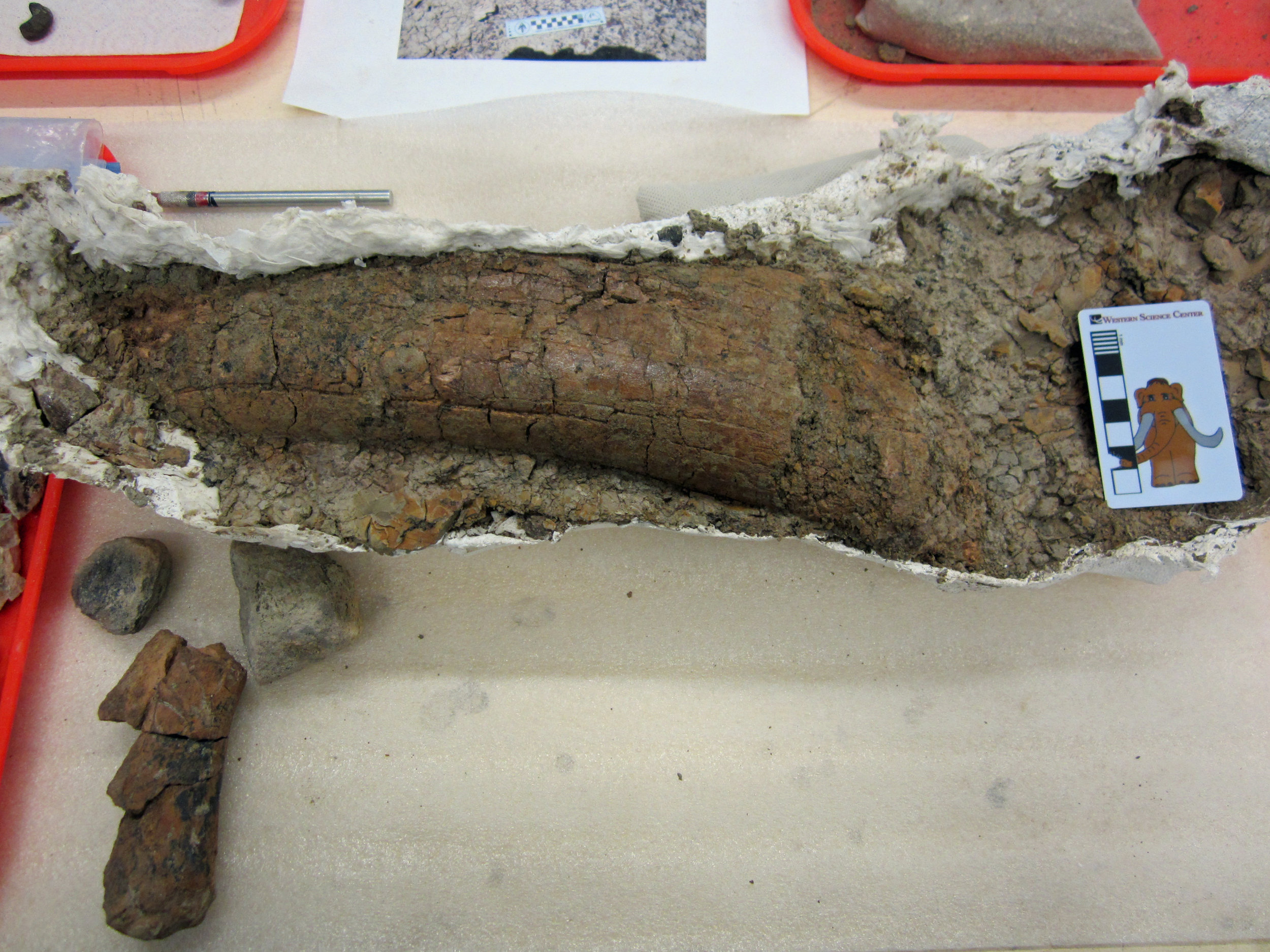
 We have discovered many isolated dinosaur limb bones in the Upper Cretaceous Menefee Formation of New Mexico. Although it's difficult to infer the full appearance of a dinosaur from a single bone, even isolated bones can tell us a great deal about what kinds of dinosaurs were roaming around 80 million years ago, and can be very surprising!This bone was collected by my colleagues at the Zuni Dinosaur Institute for Geosciences and volunteers with the Southwest Paleontological Society in 2015. We have been thinking that it was possibly two dinosaur limb bones next to each other. However, Western Science Center volunteer Joe Reavis opened up the plaster jacket this week, and it turns out that it contains a single massive shin bone, a tibia. The bone is still partially encased in mudstone, but we'll keep working on it to determine what sort of dinosaur walked around on this immensely thick bone.Post by Curator Dr. Andrew McDonald.
Fossil Friday - dinosaur limb bone
 In the Western Science Center lab, we're making steady progress through several field seasons' worth of fossils from the Menefee Formation in New Mexico. We're working on fossils of dinosaurs, crocodiles, turtles, and plants, all dating to around 80 million years ago.This week, WSC volunteer Joe Reavis began prepping a fairly hefty dinosaur limb bone that was collected in 2016 by me and colleagues from the Zuni Dinosaur Institute for Geosciences and Southwest Paleontological Society. We're not yet certain what type of dinosaur this is from, or even which bone in the skeleton this is. One of the ends of the bone had eroded out onto the hillside prior to discovery, so we need to see if we can piece it back together and reattach it to the rest of the bone. There's always more to learn!Post by Curator Dr. Andrew McDonald.
Fossil Friday - ceratopsian vertebra
 On January 25, I showed you a horned dinosaur vertebra that was still partially encased in its plaster field jacket. Western Science Center volunteer John Deleon has been working hard on this vertebra for the last several weeks and has made great progress. The vertebra is now completely freed from its jacket, and is awaiting a bit more matrix removal and gluing before it is finished.This vertebra is part of a partial ceratopsian skeleton excavated in New Mexico in 2018 by the Western Science Center, Zuni Dinosaur Institute for Geosciences, and Southwest Paleontological Society. At between 80 and 79 million years old, this skeleton is among the oldest specimens of large horned dinosaurs from North America. We are not yet certain what kind of horned dinosaur this is, but there are numerous more bones from the same animal waiting to be prepared in the WSC lab.Post by Curator Dr. Andrew McDonald.
Fossil Friday -crocodile skull
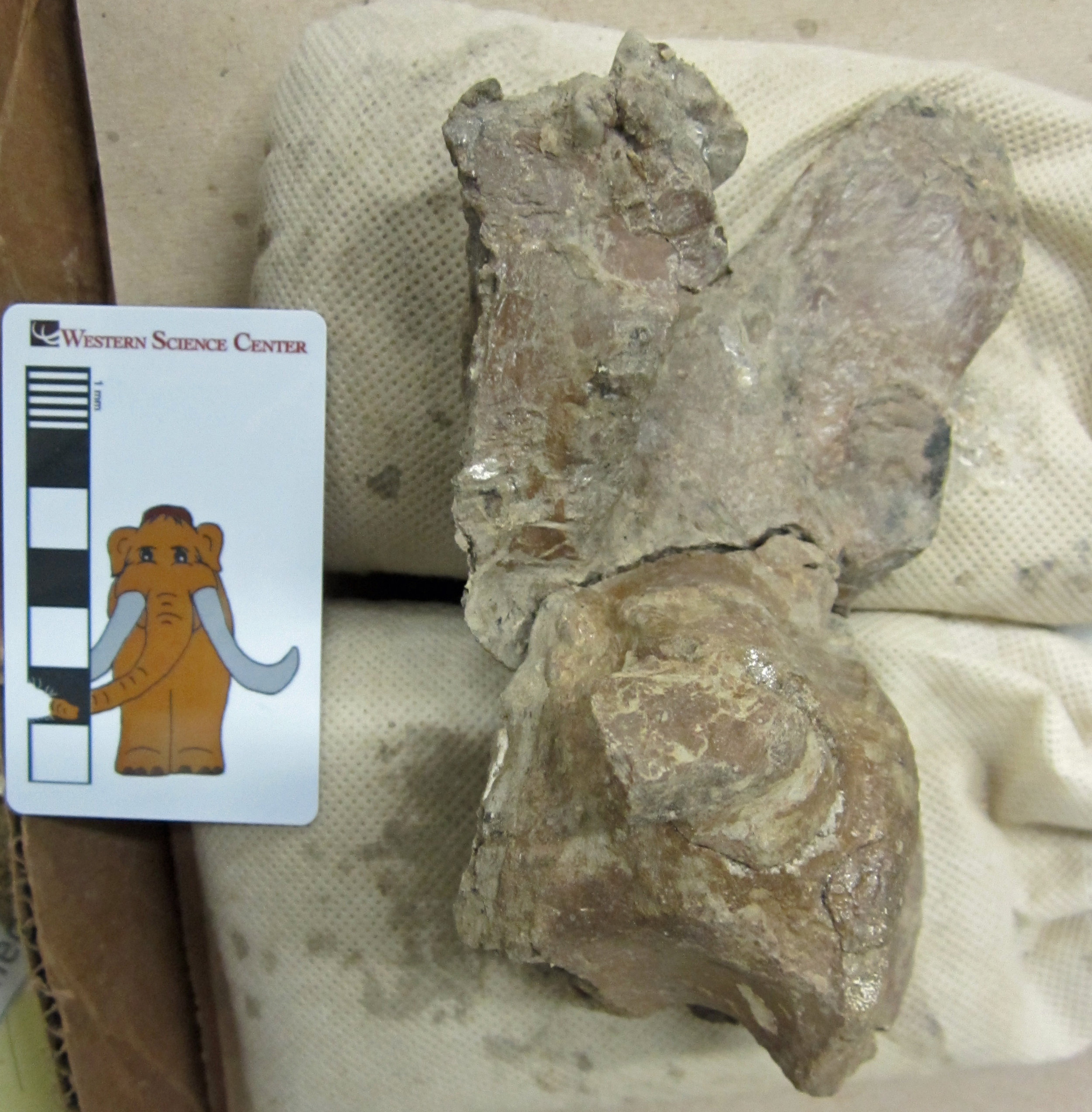
 For Fossil Friday today, I want to give you an update on one of the fossils we collected in New Mexico last year. On December 14, 2018, I posted the smashed up skull of a small crocodile from the Menefee Formation, around 79 million years old.At the time, we had just removed this little skull from its plaster jacket. Although broken apart by erosion, from the deeply pitted texture of the bones and presence of the occipital condyle (the ball at the back of the skull that articulates with the first neck vertebra), it was clear that this is indeed a small crocodile skull. In order to piece it back together, the bone fragments would need to be removed from the surrounding rock. However, it appeared that none of the fragments had moved too far from their original positions. Therefore, it was important to have a record of the condition of the fossil before preparation began.To that end, WSC Educator Brett Dooley scanned the fossil with our NextEngine laser scanner, and Director Alton Dooley created a digital 3D model. We then printed it on one of the museum's 3D printers. We also took photographs of the fossil. Using the printed replica and photos as a guide, WSC lab volunteer Joe Reavis has been teasing the bone fragments from the rock in order to clean them and fit them together. As of now, the mass of bone fragments has been disassembled, and Joe and I have been able to start fitting pieces together and identifying the bones.The bones represent much of the back end of the skull, including bones that surrounded the brain (parietal, basisphenoid, and basioccipital with the occipital condyle), bones from the adjacent skull roof (left squamosal), and the bones with which the lower jaw would have articulated (the left and right quadrates). There is much work still to do, but once finished, this little skull will be a nice addition to our knowledge of the Menefee Formation's crocodiles. We hope to be able to reconstruct this region of the skull and determine to which species this specimen belongs.Post by Curator Dr. Andrew McDonald
Fossil Friday - more on Mystic the whale
 Last summer we took delivery of Mystic, a Pliocene baleen whale from Santa Cruz County. It will take us years to fully prepare Mystic, but we have started working on it, and we've made some interesting progress.Back in July, I tentatively identified Mystic as a possible member of the right whale family (the Balaenidae), based on its large size, the arched rostrum (upper jaw), and the lack of a coronoid process on the mandible. Below is a skeleton of the modern bowhead whale, Balaena mysticetus, showing those features (photo from the Royal Belgian Institute of Natural Sciences, taken by Nick Fraser):
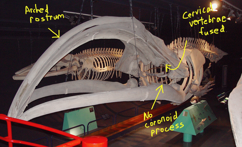 The fused cervical vertebrae are not visible in the photo above, but an interesting feature of balaenids is that all seven neck vertebrae are fused in both modern genera at a very young age, and the first six are fused in the only fossil genus with a known vertebral column, Balaenula (example below from the Pliocene of Virginia, at the North Carolina Museum of Natural Sciences):
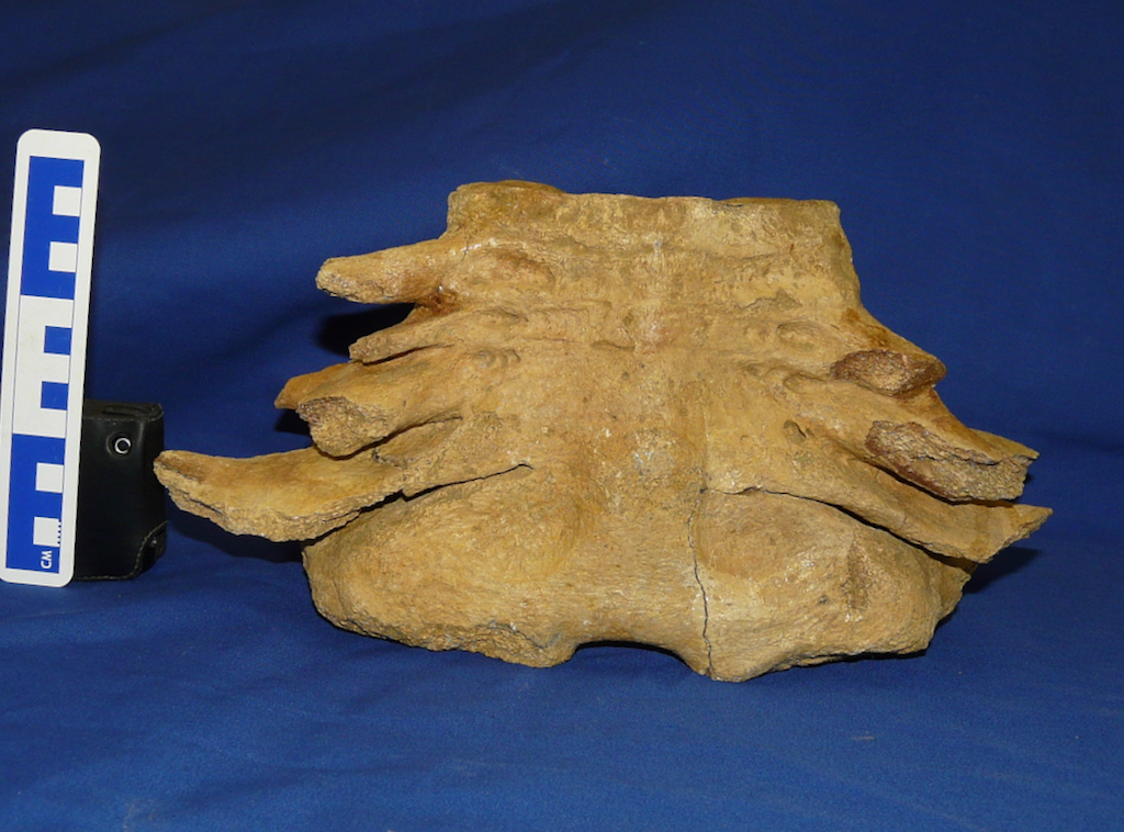 In recent weeks, we've been working around the back of Mystic's head, and noticed a series of vertebrae (after the photo is a version with the vertebrae circled):
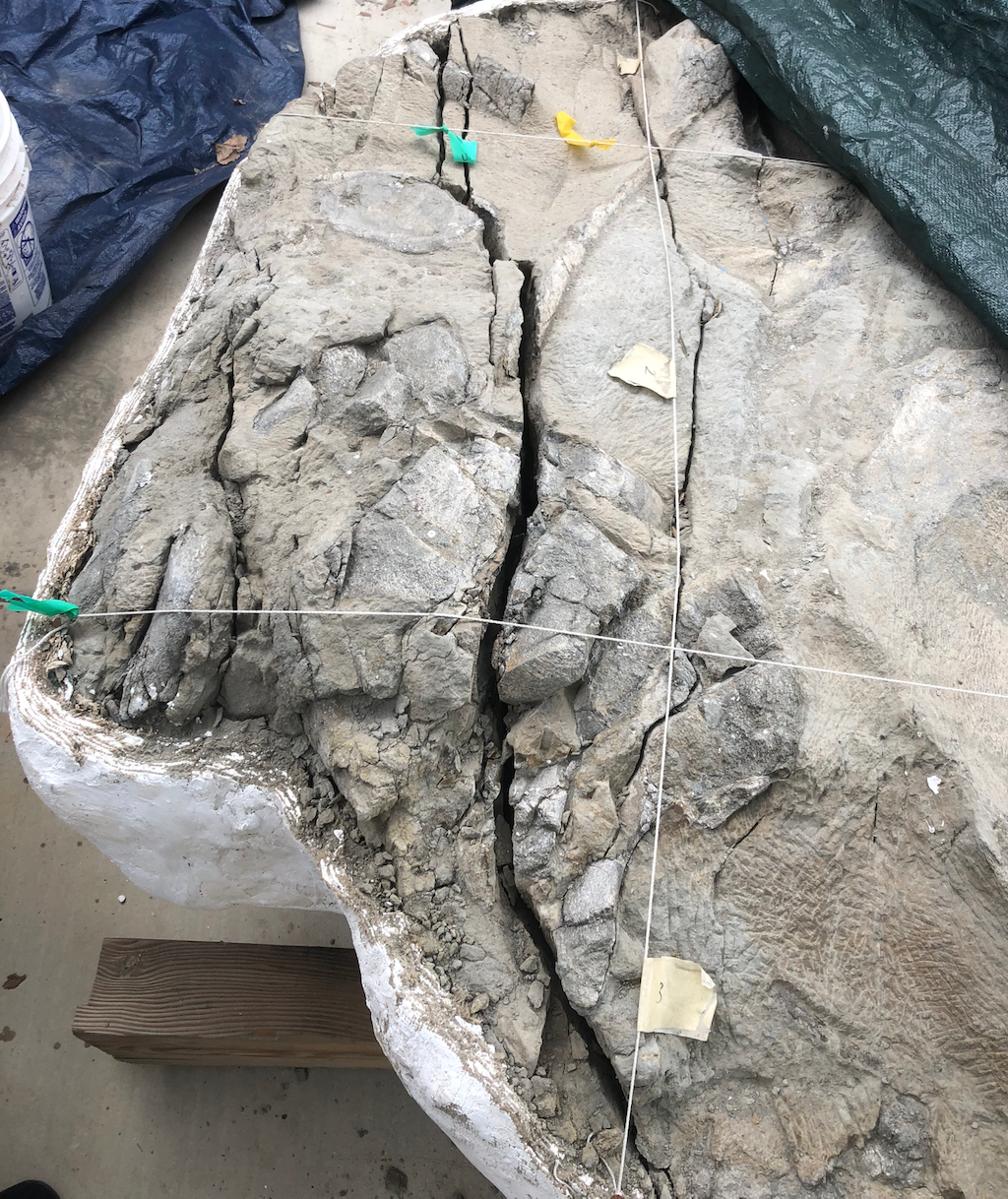
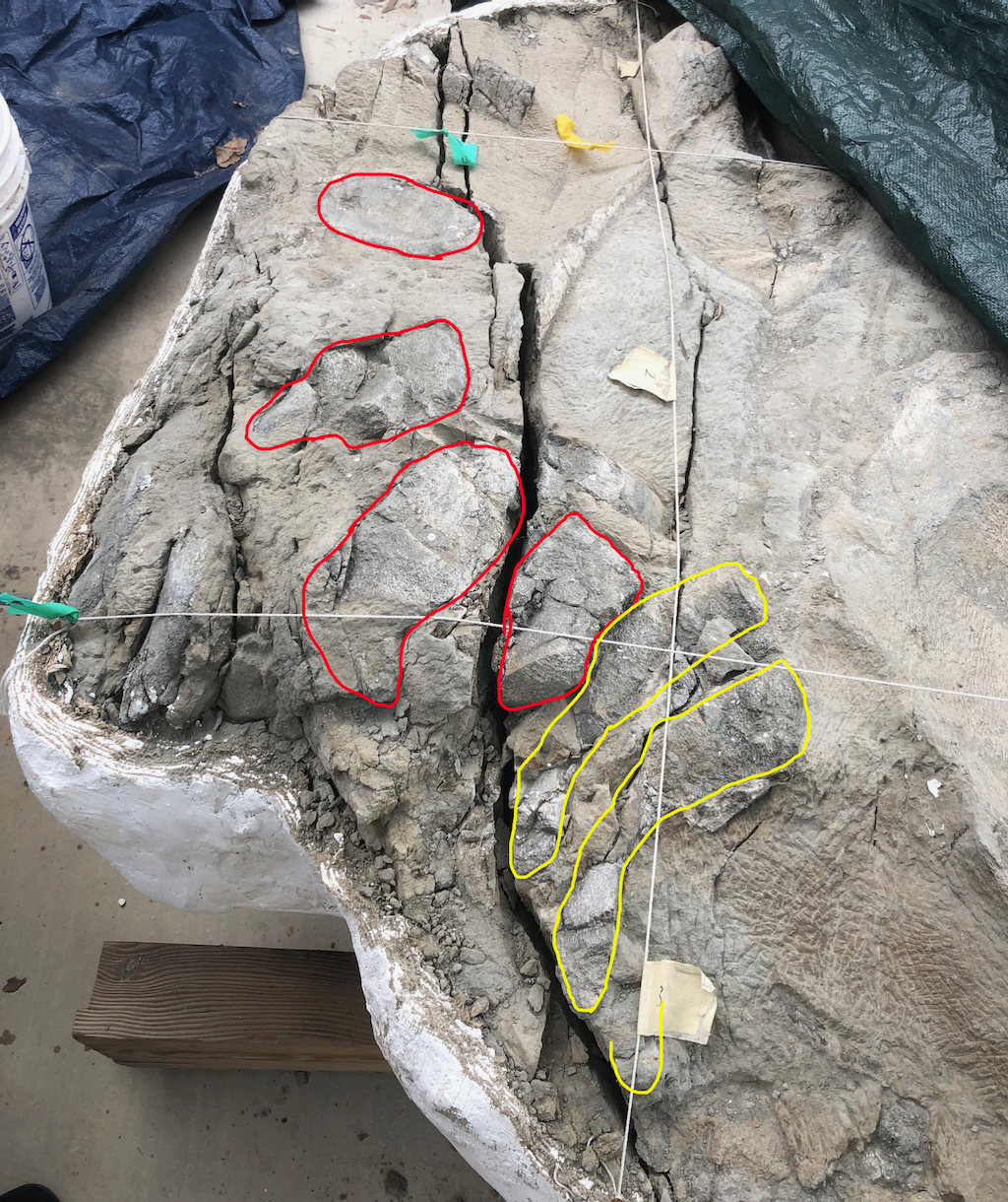 Most of these vertebrae are articulated, or nearly so. But the ones in yellow are of particular interest. They have transverse processes with no obvious rib articulations, and the vertebral centra are very short. That makes them look like cervical vertebrae, but they aren't fused. As mentioned above, all known members of the right whale family have fused cervicals. So, do we have other options?Balaenopterids (blue whales, humpback whales, and their relatives) have unfused cervical vertebrae, but their rostra are only slightly arched, and, more significantly, they all have very large, prominent coronoid processes (fin whale Balaenoptera physalis below from the Los Angeles County Museum):
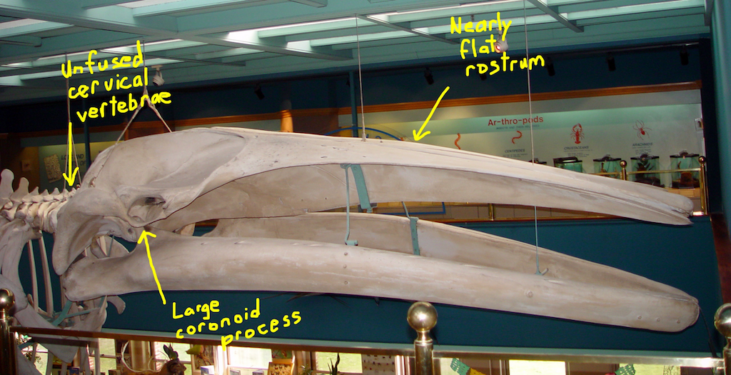 However, there are also gray whales (example below from the Monterey Bay Aquarium):
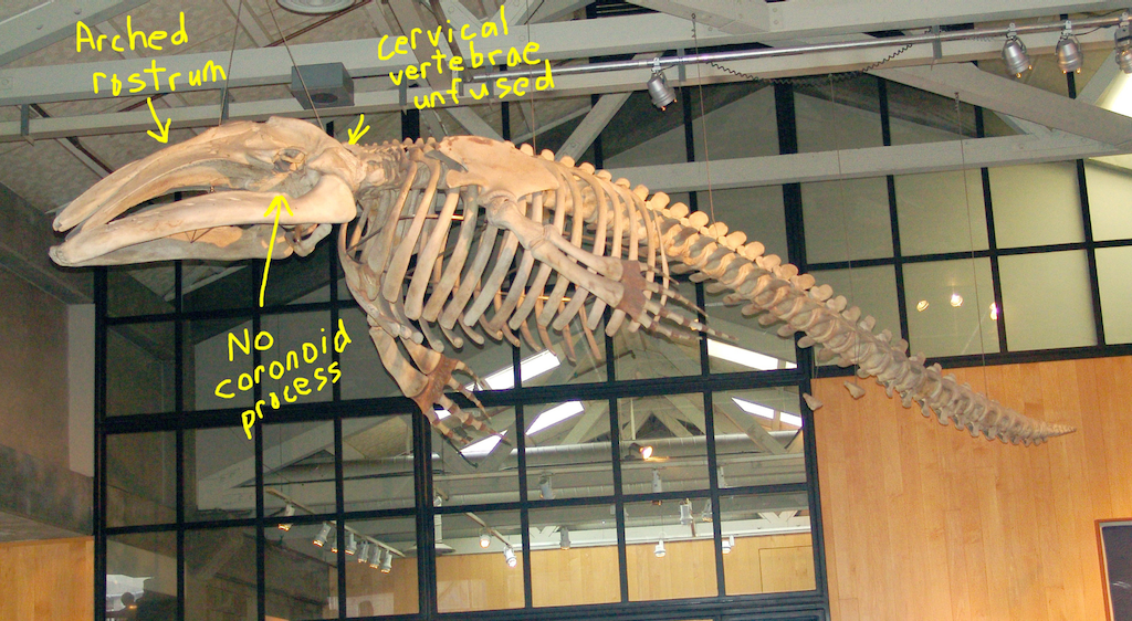 Like balaenopterids (and, in fact, all mysticetes except the right whales), gray whales have unfused cervical vertebrae. But, like right whales, they have an arched rostrum (although not as arched as in the right whales) and lack a coronoid process.If we're interpreting Mystic's features correctly, I now think there's a good chance it could be a gray whale instead of a right whale. There have been other fossil gray whales found in Pliocene deposits from California, especially from the San Diego Formation, but they don't seem to be terribly common, so this would still be a great find.Stay tuned for more over the next five years or so!
Fossil Friday - ceratopsian vertebra
 Identifying fossil bones can be quite the challenge. Fossils might be broken, scattered, or distorted from the weight of the rock encasing them. This dinosaur bone was collected in June 2018 by the Western Science Center, Zuni Dinosaur Institute for Geosciences, and Southwest Paleontological Society in the Menefee Formation of New Mexico. We know from other bones collected in the same spot that we are dealing with the 79-million-year-old partial skeleton of a ceratopsid, one of the large horned dinosaurs related to Triceratops.In the field, all we could see was a flat but somewhat intricate surface that we thought might be a skull bone. WSC volunteer John Deleon recently started prepping this fossil out of its small plaster jacket, so we can see more of its shape and features for the first time. Turns out this bone is actually a highly distorted vertebra that has been flattened side to side. Right now it is visible only from the left side. Many characteristics of a typical vertebra are observable, including the centrum (the main body of the vertebra), the neural spine, and the left prezygapophysis and postzygapophysis (along with the centrum, these structures are where the vertebra would articulate with the vertebra in front of it and the one behind it).We have many more vertebrae, ribs, and a large hip bone to prepare from this ceratopsid, so we will have much more to say about this dinosaur in the near future.
Fossil Friday - fox squirrels and vampire's disease
 This week we opened a new permanent exhibit at Western Science Center on eastern fox squirrels. This is based on research that I did in 2011-2013 in collaboration with Dr. Nancy Moncrief, Curator of Mammalogy at the Virginia Museum of Natural History (at that time I was Curator of Paleontology at VMNH). Much of the following is taken from a post I wrote for my old blog, Updates from the Paleontology Lab, on 29 February 2012.
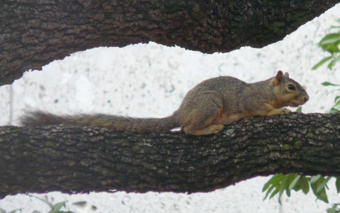 For many years, Nancy worked on the biogeographic patterns in the eastern fox squirrel, Sciurus niger (above), mostly by looking at marker genes. S. niger is an interesting species for this work because there are numerous subspecies that vary greatly in body size and coat color, as you can see in these specimens from the VMNH collections:
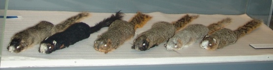 That’s a lot of variation! What makes it really interesting is that S. niger is a temperate species that doesn’t tolerate cold weather very well (they don’t hibernate). That means that their range was probably very restricted in the Pleistocene during the last glacial maximum, suggesting that all that phenotypic diversity may have arisen in less than 10,000 years.I was not involved in Nancy’s research, but because I do some biogeographic work Nancy would occasionally ask me to read her manuscripts or listen to her lectures to provide feedback from a paleontologist’s perspective. As a result I was somewhat familiar with her work.Then, in 2009 I was asked to write a book chapter reviewing the published record of vertebrate fossils in Virginia (which was finally published in 2016). While tabulating these species I noticed that, while the genus Sciurus had been reported from the Virginia Pleistocene, there were no reports of S. niger. Almost all the records were listed as Sciurus sp.Realizing that being able to tell when fox squirrels show up in different places could be useful for Nancy’s work, I asked her about the lack of definitive species-level identifications. The problem, she explained, is that it’s really difficult to distinguish S. niger from the closely related, sympatric, eastern gray squirrel (Sciurus carolinensis, below) when only the skeleton is available.
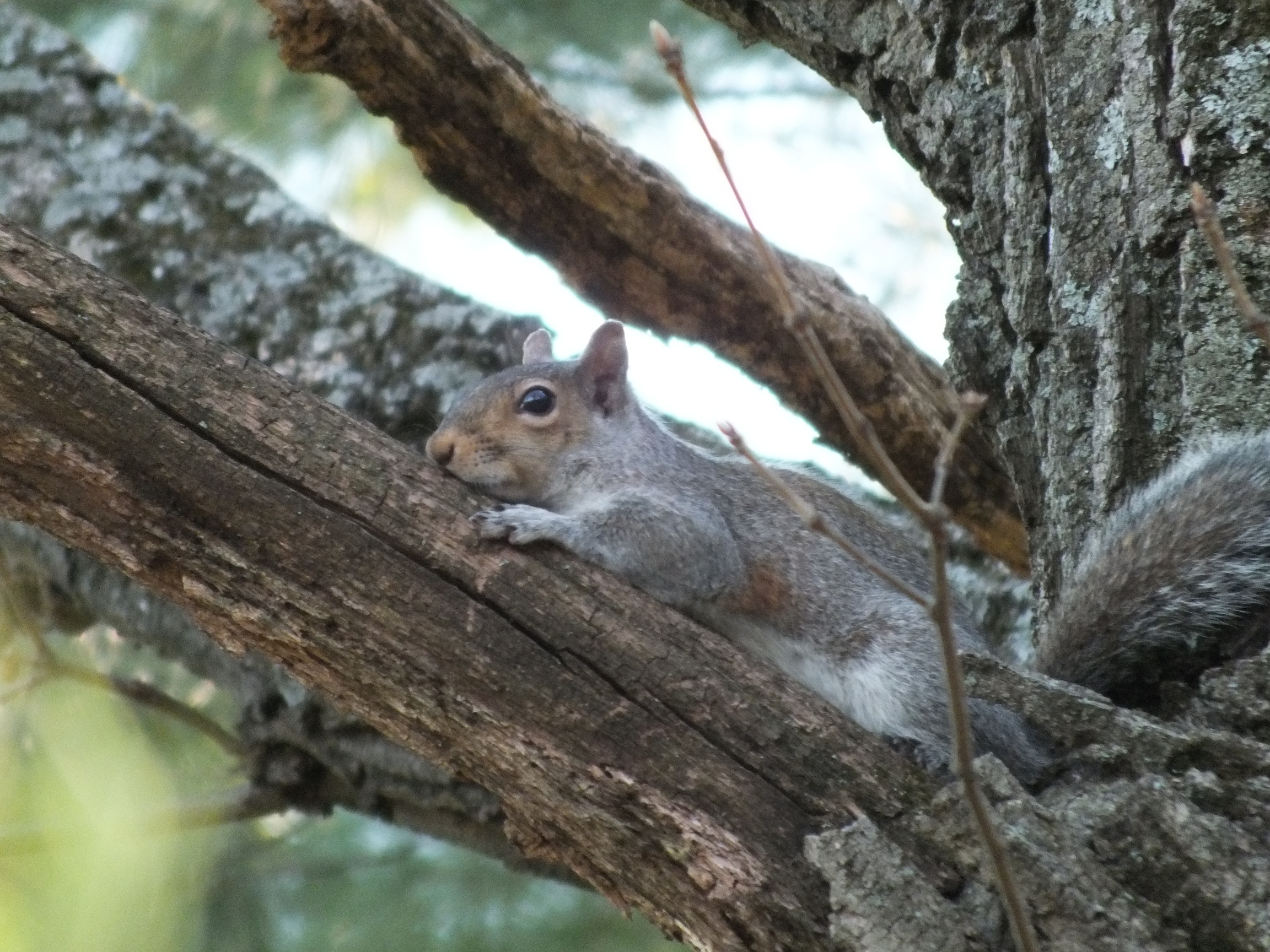 The problem is that tree squirrels are pretty conservative in their skeletal morphology, especially in the postcranial skeleton, with few changes since the Miocene at least. The average size and maximum size of fox squirrels are greater than in gray squirrels. However, there is considerable overlap; the largest gray squirrels are bigger than the smallest fox squirrels. So using body size for identification only works for really big specimens.There are also some detail differences in the skull and mandible, but they tend to be somewhat inconsistent. Easily the most reliable feature is the presence of a rudimentary upper 3rd premolar in S. carolinensis, which is absent in S. niger. Below is the upper right tooth row of S. carolinensis, top, and S. niger, bottom. In each specimen anterior is to the right. Note the tiny, peg-like premolar at the front of the tooth row in S. carolinensis only:
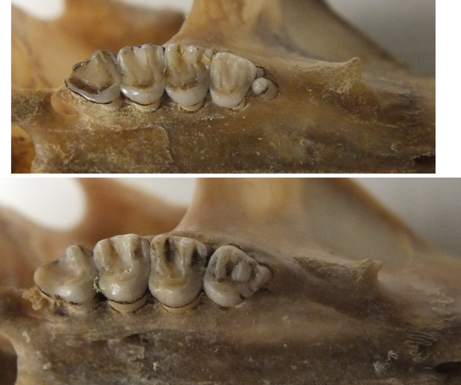 Unfortunately, it’s pretty rare to preserve that particular part of the maxilla, so the extra premolar is of limited use in identifying isolated bone fragments in Pleistocene deposits. In an irrational fit of optimism, I suggested to Nancy that I might be able to come up with features for distinguishing between the two species based on isolated material, especially since the VMNH collections include hundreds of modern squirrel skeletons to use as references.After several frustrating days looking at squirrel bones, I was ready to give up. There simply don’t seem to be any consistent features for distinguishing postcranial remains other than size, which is unreliable. Then, as Nancy and I sat together discussing squirrel trivia, she asked if I knew that fox squirrel bones glow pink under ultraviolet light. It hit us both at the same time; only fox squirrel bones fluoresce under UV light! Here’s an S. niger skull under visible light (left), and the same skull under UV light (right):
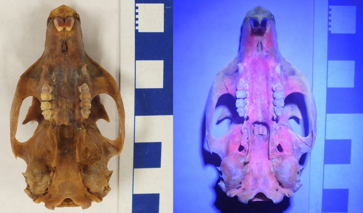 So here’s the deal: eastern fox squirrels almost universally have a genetic condition called congenital erythropoietic porphyria (CEP). This is caused by a mutation in one of the genes involved in the heme chain (heme is a key component of hemoglobin). Specifically, animals with CEP can’t properly metabolize the enzyme uroporphyrin, which as a result starts to build up in the urine, bones, and teeth. Uroporphyrin fluoresces pink when exposed to ultraviolet light.As I mentioned above, CEP is normally not present in gray squirrels, so we can potentially use the presence of fluorescence to distinguish between fox and gray squirrels, even when there’s a size overlap. For example, the skulls below are organized by increasing size from left to right. Which ones are S. niger, and which ones are S. carolinensis?
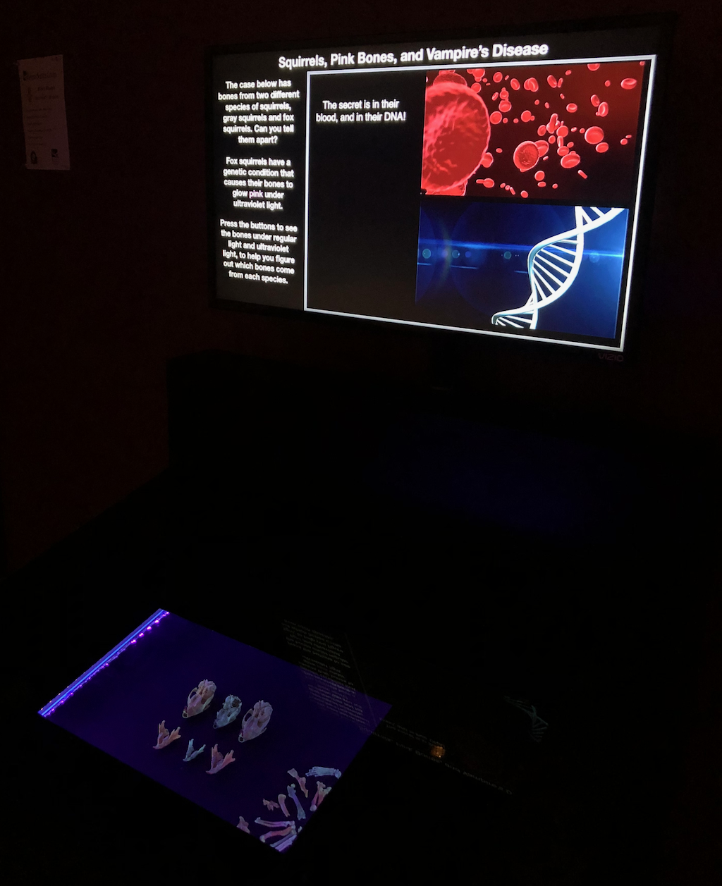
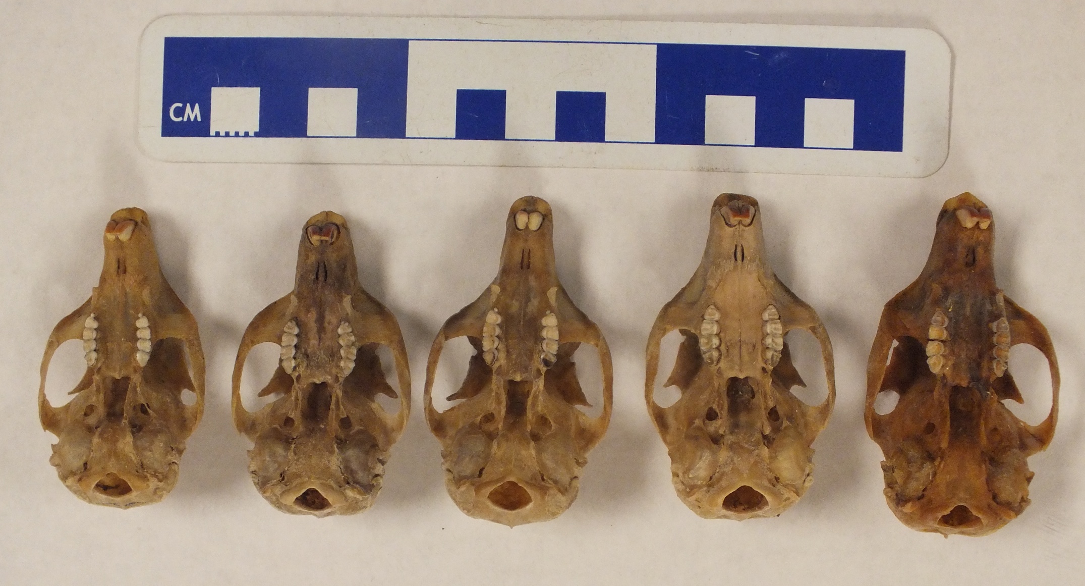 Here are the same skulls under UV light:
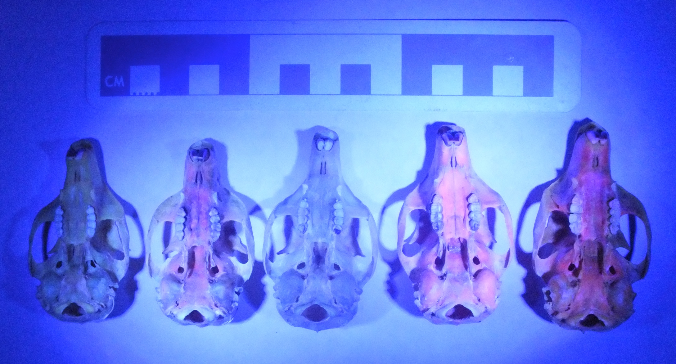 The 2nd, 4th, and 5th skulls all fluoresce, and are all fox squirrels.CEP has been reported as a rare condition in several different mammal species, including humans. It’s typically a pretty dreadful condition that results in photosensitivity, skin lesions, and anemia among other problems. The combination of sensitivity to sunlight and anemia (requiring blood transfusions) has led to CEP sometimes being called "Vampire's disease". For some unknown reason, eastern fox squirrels don’t seem to suffer from the adverse effects of CEP even though they have elevated uroporphyrin levels.Biologists have known since the 1930’s that fox squirrels have fluorescent bones (Turner, 1937), and that sometimes they’re even pink under visible light. There were a few studies in the 1970’s looking at the condition in some detail (Levin and Flyger 1971, for example), but somehow this knowledge was never applied to fossil remains. The question, of course, is that since a particular protein is responsible for the fluorescence, would it still be detectable in ancient specimens?The Florida Museum of Natural History lists several specimens of S. niger among their Pleistocene/early Holocene collections, and they happened to be the host for the 2011 SeAVP meeting. The day before the meeting I spent a few hours in their collection with a tray of squirrel bones from Devil’s Den Sinkhole and a UV light. Sure enough, even though these bones have been sitting at the bottom of a sinkhole (that’s currently flooded) for at least 7,000 years*, several of the bones still fluoresce. The best example was this partial skull:
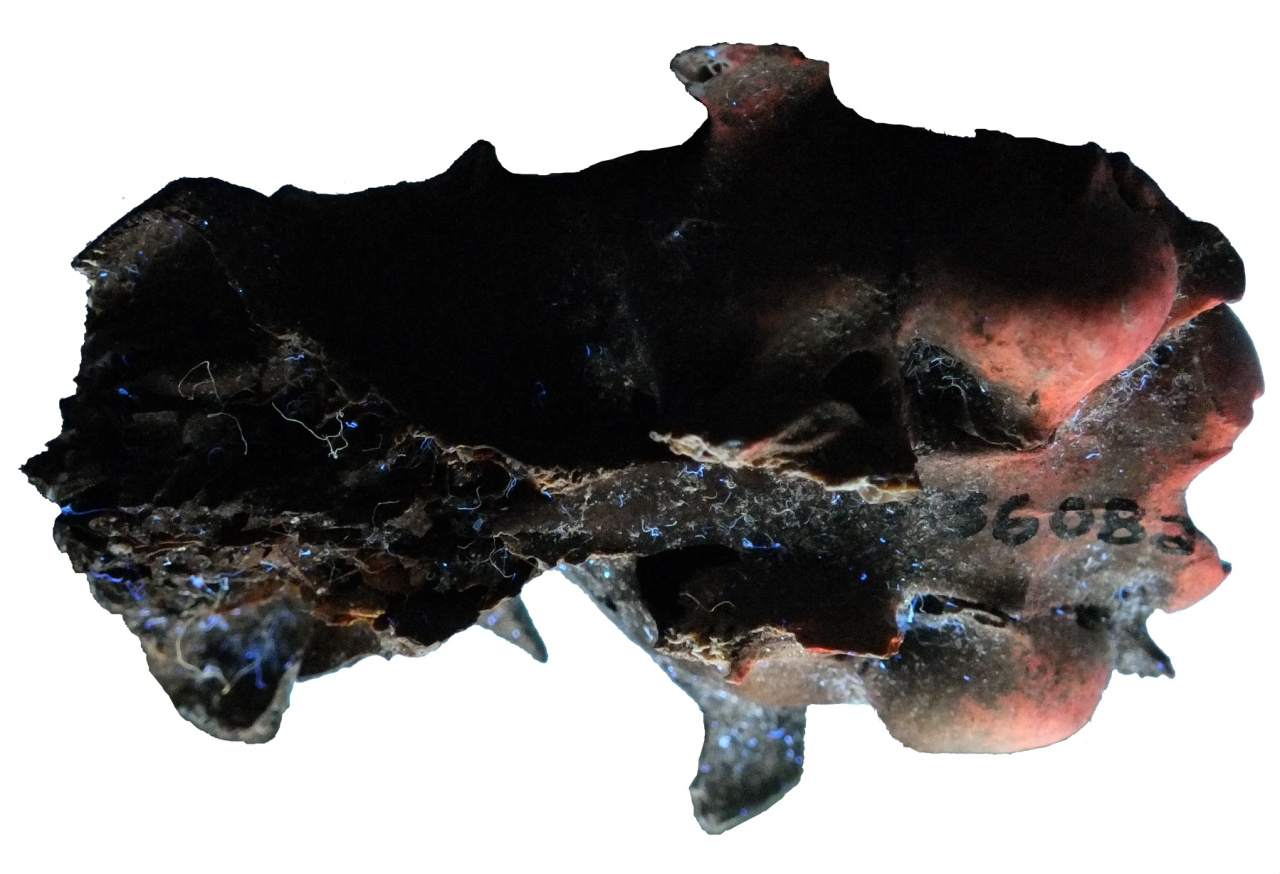 There’s definitely a high “coolness” factor here; we diagnosed a genetic condition in a late Pleistocene/early Holocene squirrel! But in addition to that, we’ve given ourselves a pretty powerful tool for examining ancient squirrels. To be sure, there are limitations: the fluorescence seems to disappear if the bones have been mineralized, and the condition may not be truly universal in S. niger. Therefore, this only works as a positive test for the presence of S. niger; the absence of fluorescence doesn’t definitively tell us anything. But on the other hand, this allows us to identify even fragmentary postcranial remains, the test is non-destructive and inexpensive, and the results are immediate. And it’s spectacularly easy to use on mixed bone deposits, including samples obtained by screening. Here’s a sample of bones from multiple taxa obtained by screening a sediment sample from a late Holocene site:
 And here’s the same sample under UV:
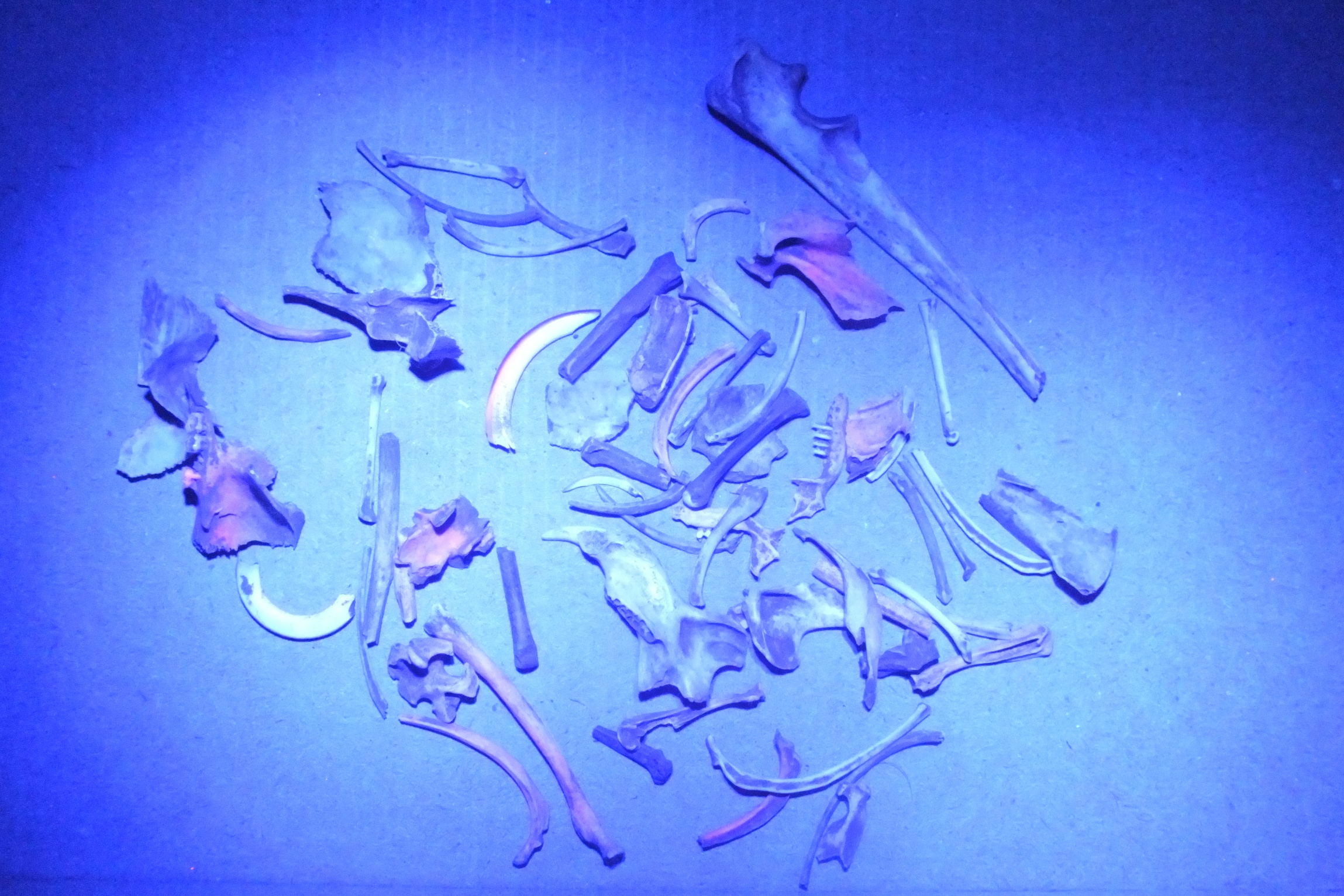 A dozen of these bones turned out to be fox squirrel (some of them are hard to spot in a photograph). They include difficult-to-identify elements like ribs; notice the two at the bottom center of the photo.Nancy and I published the results of our study of the Devil's Den fossil squirrels in 2012. In a follow-up paper in 2013, we examined squirrel remains from Native American archaeological sites in Virginia that were roughly 1,000 years old. Even though these specimens had in most cases been burned, they still fluoresced, as with this tibia:
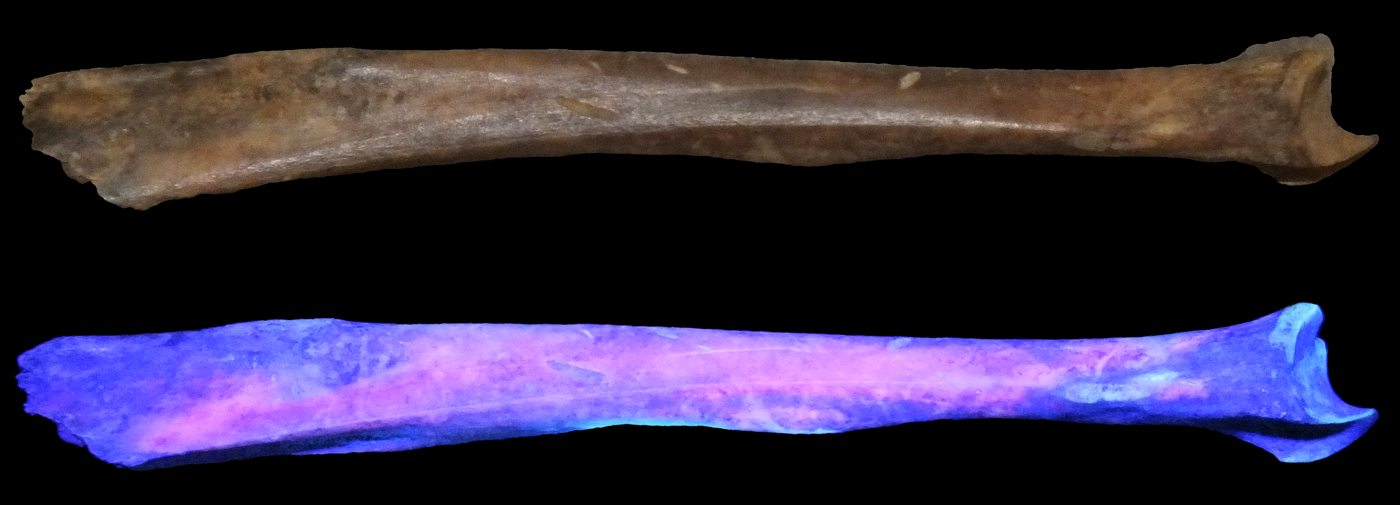 This research brought up a lot of additional questions that we never fully addressed. We found, both in the literature and from our own observations, that not all fox squirrels fluoresce equally, and a minority don’t seem to fluoresce at all. Why? Are some populations more prone to CEP, is there an ontogenetic component to uroporphyrin levels, or is there just a significant amount of individual variation? Moreover, certain elements seem to fluoresce particularly well (for example, the maxillae, the dentaries, the incisors, and the 4th upper premolars) while others often don’t fluoresce at all (like the nasals and the molars). Since moving to California, I've also learned that there is an introduced population of Eastern fox squirrels in Los Angeles County, which have been isolated from other S. niger populations now for toughly 100 years. I've not yet had the chance to examine one of these squirrels, but I wonder if their bones still fluoresce?*There’s some uncertainty about the age of the Devil’s Den deposits. They were published as being approximately 7,000 years old, but the presence of extinct animals such as mastodonts in the same deposit indicates that it is more likely late Pleistocene (Martin and Webb, 1974).References:
Dooley, Alton C., Jr., and Nancy D. Moncrief, 2012. Fluorescence Provides Evidence of Congenital Erythropoietic Porphyria in 7,000-Year- Old Specimens of the Eastern Fox Squirrel (Sciurus niger) from the Devil’s Den. Journal of Vertebrate Paleontology 32:495–497.Levin, Ephraim Y., and Vagn Flyger, 1971. Uroporphyrinogen III Cosynthetase Activity in the Fox Squirrel (Sciurus niger). Science 174:59–60.
Martin, R. A., and S. D. Webb. 1974. Late Pleistocene mammals from the Devil’s Den fauna, Levy County; pp. 114–145 in S. D. Webb (ed.), Pleistocene Mammals of Florida. University of Florida, Gainesville, Florida.
Moncrief, Nancy D., and Alton C. Dooley, Jr., 2013. Using fluorescence of bones and teeth to detect remains of the Eastern fox squirrel (Sciurus niger) in archaeological deposits. Southeastern Archaeology 32:46-53.Turner, William J., 1937. Studies on Porphyria. I. Observations on the Fox-squirrel, Sciurus niger. Journal of Biological Chemistry 118:519–530.
Fossil Friday - hadrosaur metatarsal
 Discovering a dinosaur skeleton is a rare and special event.Most often, when out exploring in the badlands of New Mexico, we find isolated bones. However, even an isolated bone offers a wealth of information, about what kinds of animals were living in the area 79 million years ago, the depositional environment, and any unusual anatomical features or pathologies. Every summer, we collect numerous isolated bones of dinosaurs, crocodiles, and other animals from the Menefee Formation.Western Science Center prep lab volunteer John Deleon recently began preparing this dinosaur bone, which was collected by Southwest Paleontological Society volunteers Ben Mohler and Jake Kudlinski during our expedition last June. No other bones were found nearby, but this bone itself is well preserved and certainly worthy of the effort and study. It is a metatarsal, one of the large foot bones situated between the ankle and the toes, and probably belonged to a duck-billed plant-eating hadrosaur. Isolated hadrosaur bones, mostly limb bones and vertebrae, are the most commonly found dinosaur fossils in the Menefee Formation, suggesting that hadrosaurs probably were quite abundant in the ancient ecosystem. We'll be able to compare this bone to other limb bones we have collected over the years.Post by Curator Dr. Andrew McDonald.
Fossil Friday - Zygolophodon tooth
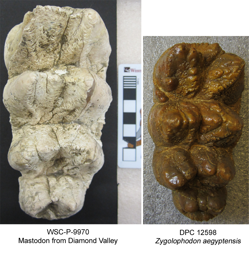 Last week, I visited the Division of Fossil Primates at the Duke Lemur Center in Durham, North Carolina. They have a great collection of fossil proboscideans from Egypt, including early forms like Moeritherium and Paleomastodon, as well as later, larger species such as Gomphotherium angustidens.I was there especially to see four teeth, the only known fossils of Zygolophodon aegyptensis. The genus Zygolophodon is long-lived and widespread, including many other species from Europe, Asia, and North America. At about 18 million years old, Z. aegyptensis is among the oldest. Zygolophodon is a member of the group Mammutidae, making it a close relative of North America's mastodons, including those found at Diamond Valley and housed at Western Science Center.The image here is DPC 12598, an upper molar (right M3) of Zygolophodon aegyptensis, and a left M3 of a mastodon from Diamond Valley (WSC-P-9970) set to the same scale. My Western Science Center colleagues and I are working with paleontologists at several other institutions to explore the whole evolutionary history of Mammutidae. Visiting other museums to examine fossils of other species and comparing them to the bounty of mastodons from Diamond Valley is one of the first steps on this journey.My thanks to Catherine Riddle of the Division of Fossil Primates for her assistance during my visit.Post by Curator Dr. Andrew McDonald.
Fossil Friday - grasshopper mouse
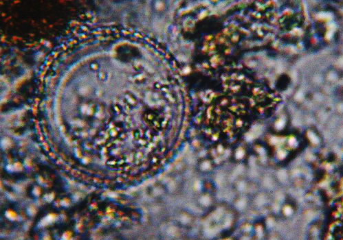
 Abstract deadlines are coming up for conferences taking place in the first 6 months of 2019, so we're trying to collect as much data as possible for our submissions. One of these is a follow-up to our abstracts from last year on the Harveston locality in Temecula, but this time looking at the small animals ("microvertebrates"). One of our high school volunteers (or "Max's Minions", as we call them), Charlotte Hohman, is taking the lead on this project, sorting vials of tiny rodent, lizard, and bird bones and trying to identify them. This week we may have found a bone from a relatively uncommon mouse, the most metal mouse in Southern California.The bone in question is a left calcaneus (heel bone). The image isn't the greatest because it was taken by holding an iPhone up to a microscope eyepiece; we just wanted a quick reference image. The total length is about 5 mm.After comparing this to references, we're pretty sure this calcaneus comes from a southern grasshopper mouse, Onychomys torridus. If correct, this is our first grasshopper mouse from Harveston. Onychomys is known from the Diamond Valley Lake fauna, but is quite rare compared to the other rodents (in fact, I think we have more mastodons than grasshopper mice!).So this is just a mouse; what makes it metal? Unlike their close relatives such as deer mice, which are omnivores leaning toward herbivory, grasshopper mice are voracious predators. They primarily eat insects and other arthropods, but will eat other mice on occasion.They are known especially for eating bark scorpions. Apparently, when stung by a scorpion, the mouse's pain receptors shut down; even though they may get stung multiple times, they don't feel the pain. Northern grasshopper mice will attack centipedes, which are also venomous; the mouse is just fast and sting (I haven't been able to confirm if southern grasshopper mice also eat centipedes).And, if all that isn't enough, grasshopper mice are territorial. But their territories can be over 20 acres (!), and they'll warn away interlopers by howling with a high-pitched scream. Presumably if that doesn't work they just eat the invader.